. 2000 Aug;20(15):5516-28.
doi: 10.1128/MCB.20.15.5516-5528.2000.

Bop1 is a mouse WD40 repeat nucleolar protein involved in 28S and 5. 8S RRNA processing and 60S ribosome biogenesis

Affiliations

Bop1 is a mouse WD40 repeat nucleolar protein involved in 28S and 5. 8S RRNA processing and 60S ribosome biogenesis

Z Strezoska et al. Mol Cell Biol. 2000 Aug.

Abstract

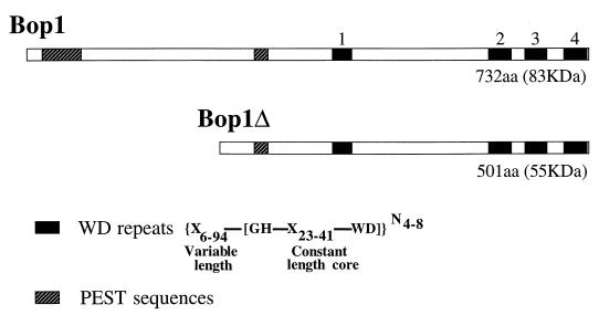
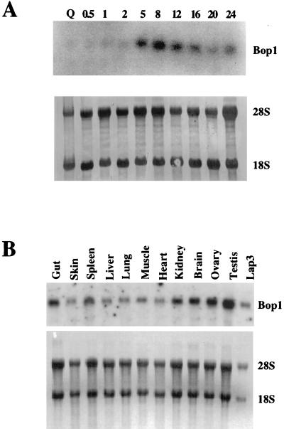
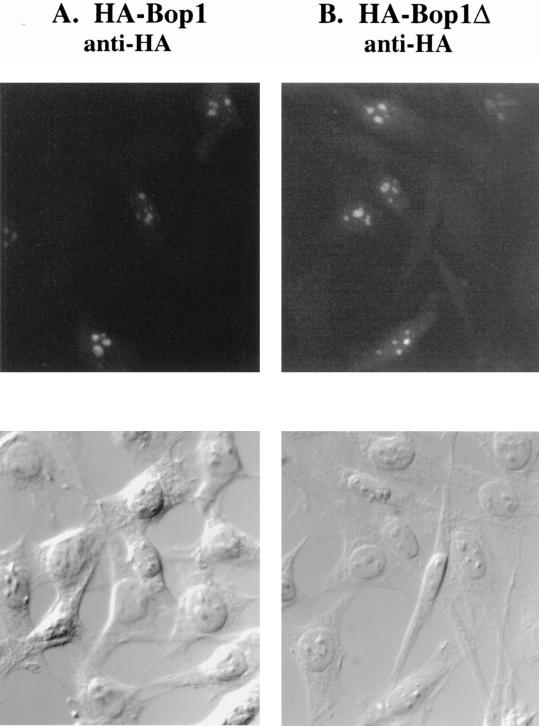
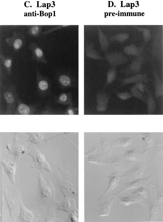
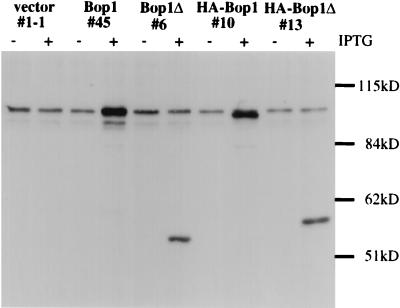
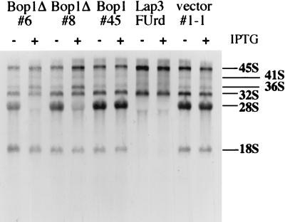
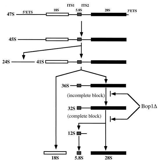
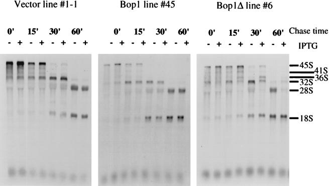
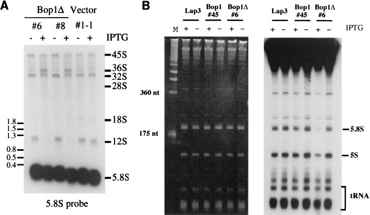
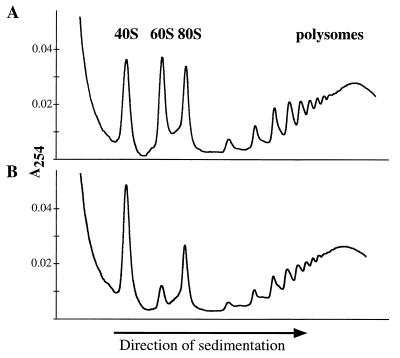
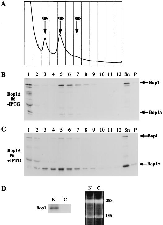
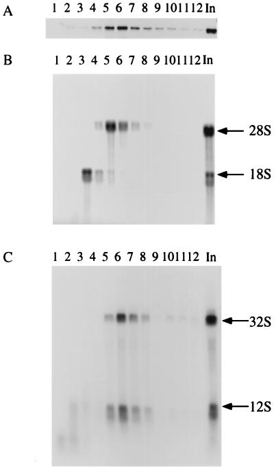
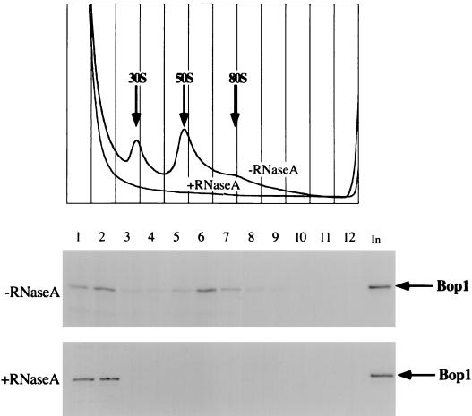
We have identified and characterized a novel mouse protein, Bop1, which contains WD40 repeats and is highly conserved through evolution. bop1 is ubiquitously expressed in all mouse tissues examined and is upregulated during mid-G(1) in serum-stimulated fibroblasts. Immunofluorescence analysis shows that Bop1 is localized predominantly to the nucleolus. In sucrose density gradients, Bop1 from nuclear extracts cosediments with the 50S-80S ribonucleoprotein particles that contain the 32S rRNA precursor. RNase A treatment disrupts these particles and releases Bop1 into a low-molecular-weight fraction. A mutant form of Bop1, Bop1Delta, which lacks 231 amino acids in the N- terminus, is colocalized with wild-type Bop1 in the nucleolus and in ribonucleoprotein complexes. Expression of Bop1Delta leads to cell growth arrest in the G(1) phase and results in a specific inhibition of the synthesis of the 28S and 5.8S rRNAs without affecting 18S rRNA formation. Pulse-chase analyses show that Bop1Delta expression results in a partial inhibition in the conversion of the 36S to the 32S pre-rRNA and a complete inhibition of the processing of the 32S pre-rRNA to form the mature 28S and 5.8S rRNAs. Concomitant with these defects in rRNA processing, expression of Bop1Delta in mouse cells leads to a deficit in the cytosolic 60S ribosomal subunits. These studies thus identify Bop1 as a novel, nonribosomal mammalian protein that plays a key role in the formation of the mature 28S and 5.8S rRNAs and in the biogenesis of the 60S ribosomal subunit.


Figures

FIG. 1
FIG. 1
Structural features of the Bop1 protein. Schematic representation of the full-length murine Bop1 protein (732 aa) and the amino-terminally truncated Bop1Δ (501 aa). The four WD repeats, whose consensus structure is as indicated, are shown as solid boxes. Repeats 1 and 4 are close to the consensus structure, while repeats 2 and 3 are more divergent. PEST sequences, often associated with short-lived regulatory proteins, are shown by hatched boxes.
FIG. 2
FIG. 2
Expression of bop1. (A) BALB/c 3T3 cells were brought to quiescence (Q) by serum starvation and restimulated with 10% fetal bovine serum. RNA was isolated at the indicated times (in hours) after serum stimulation and analyzed by Northern blotting with 32P-labeled bop1 cDNA as probe. The RNA blot was stained with methylene blue (lower panel) to show the relative amounts of rRNAs, indicating equal loading of the samples. (B) RNA was isolated from different adult mouse tissues and analyzed by Northern blotting with labeled bop1 cDNA as probe. The lower panel shows methylene blue staining of the same blot to control for loading of the RNA.
FIG. 3
FIG. 3
Subcellular localization of Bop1 by indirect immunofluorescence. (A and B) Pools of LAP3 cells stably transfected with vectors that express HA-tagged Bop1 (A) or Bop1Δ (B) were grown on coverslips, induced with IPTG for 12 h, fixed, permeabilized, and stained with monoclonal anti-HA antibody. Antibody-antigen complexes were detected with FITC-conjugated anti-mouse antibody and visualized by fluorescence microscopy. An image of the same field visualized with DIC optics is shown in the lower panels. (C and D) To localize the endogenous Bop1 protein, LAP3 cells were grown on coverslips, fixed, permeabilized, and stained with affinity-purified, polyclonal anti-Bop1 antibodies (C) or the preimmune serum as a control (D).
FIG. 3
FIG. 3
Subcellular localization of Bop1 by indirect immunofluorescence. (A and B) Pools of LAP3 cells stably transfected with vectors that express HA-tagged Bop1 (A) or Bop1Δ (B) were grown on coverslips, induced with IPTG for 12 h, fixed, permeabilized, and stained with monoclonal anti-HA antibody. Antibody-antigen complexes were detected with FITC-conjugated anti-mouse antibody and visualized by fluorescence microscopy. An image of the same field visualized with DIC optics is shown in the lower panels. (C and D) To localize the endogenous Bop1 protein, LAP3 cells were grown on coverslips, fixed, permeabilized, and stained with affinity-purified, polyclonal anti-Bop1 antibodies (C) or the preimmune serum as a control (D).
FIG. 4
FIG. 4
Characterization of the affinity-purified anti-Bop1 antibodies. Clonal lines of LAP3 cells stably transfected with either the empty vector pX11 (line 1-1), pX11-Bop1 (line 45), pX11-Bop1Δ (line 6), pX11-HA-Bop1 (line 10), or pX11-HA-Bop1Δ (line 13) were either treated with 1 mM IPTG (+) for 12 h or left untreated (−). Cells were lysed in RIPA buffer, and equal amounts of protein from each sample were resolved by SDS-PAGE (8% polyacrylamide) and transferred to a nitrocellulose filter. The blot was incubated with affinity-purified anti-Bop1 antibodies, and chemiluminescent reagents were used for detection. The affinity-purified antibodies recognized a high-molecular-mass band of ∼100 kDa (larger than the expected size of 83 kDa), which was increased significantly in a Bop1-overexpressing cell line (line 45). The predicted 55-kDa Bop1Δ fragment was detected in line 6, which inducibly expresses Bop1Δ. Addition of the HA tag caused a minor shift in the mobility of both Bop1 and Bop1Δ in clonal lines 10 and 13, respectively.
FIG. 5
FIG. 5
Expression of Bop1Δ inhibits the generation of 28S rRNA. Clonal cell lines derived from transfection of LAP3 cells with pX11-Bop1Δ (lines 6 and 8), pX11-Bop1 (line 45), or the pX11 vector (line 1-1) were either treated with IPTG (+) for 16 h or left untreated (−). Thereafter, cells were metabolically labeled with [3H]uridine (2.5 μCi/ml) for 30 min and chased in nonradioactive medium for 2 h. In other samples, LAP3 cells were treated with FUrd for 15 min prior to chase. RNA was then isolated, and equal counts per sample were electrophoresed on 1% agarose gel, transferred to a nylon membrane, and visualized by fluorography.
FIG. 6
FIG. 6
Schematic representation of the mammalian rRNA-processing pathway. Mammalian rRNA is transcribed as a single precursor which is further processed by successive nucleolytic cleavages that lead to elimination of the external transcribed spacers, 5′ETS and 3′ ETS, and the internal transcribed spacers, ITS1 and ITS2. The sedimentation coefficients (S) of various intermediates and mature products of the processing pathway are indicated. The mammalian 47S precursor is rapidly cleaved at the 5′ETS and at the 3′ETS to give rise to the 45S pre-rRNA. Further processing at the 5′ETS takes place, giving rise to a 41S rRNA precursor, which is rapidly processed to the 18S rRNA and a 36S precursor RNA that contains the sequences of the 5.8S and 28S rRNAs with an intervening sequence (ITS2). The 36S precursor then undergoes cleavage at the 5′ end to give rise to a 32S precursor, which is processed to the 28S rRNA and a 12S RNA; the 12S RNA is then further processed to form the 5.8S rRNA.
FIG. 7
FIG. 7
Bop1Δ inhibits processing of the 36S and 32S precursors to form the 28S rRNA. Clonal LAP3 cell lines transfected with either the empty pX11 vector (line 1-1), pX11-Bop1 (line 45), or pX11-Bop1Δ (line 6) were either left untreated (−) or treated with 1 mM IPTG for 16 h (+) and pulse-labeled with 3H-labeled methyl methionine for 30 min. After a chase in nonradioactive medium plus excess methionine for the indicated times, RNA was isolated, resolved on a 1% agarose gel, transferred to a membrane, and visualized by fluorography.
FIG. 8
FIG. 8
Bop1Δ expression inhibits generation of the 12S precursor and the 5.8S rRNA. (A) Clonal LAP3 cell lines transfected with either the empty pX11 vector or pX11-Bop1Δ (lines 6 and 8) were either left untreated or treated with IPTG for 16 h. RNA isolated from the same number of cells was separated on a 1% agarose gel, transferred to a nylon membrane, and hybridized with an oligonucleotide probe complementary to a region in 5.8S rRNA. (B) The parental LAP3 cells or clonal cell lines transfected with either pX11-Bop1 (line 45) or pX11-Bop1Δ (line 6) were either left untreated or treated with IPTG for 16 h and metabolically labeled with 32Pi (20 μCi/ml) for 1 h. Following a chase in nonradioactive medium for 1.5 h, RNA was isolated, and equal amounts of RNA from each sample were resolved on a 10% denaturing polyacrylamide gel, which was stained with ethidium bromide for photography (left panel) and dried for autoradiography (right panel).
FIG. 9
FIG. 9
Expression of Bop1Δ causes a deficit in 60S ribosomal subunits. The inducible Bop1Δ expression cell line, line 6, was grown in the absence (A) or presence (B) of IPTG as indicated for 38 h. Cytoplasmic extracts were isolated and separated on a 10 to 45% sucrose density gradient. Profiles of absorbance at 254 nm (A254) profiles revealed the positions and relative amounts of the ribosomal subunits in the gradient.
FIG. 10
FIG. 10
Bop1 and Bop1Δ cofractionate with the 50S-80S pre-RNP particles in nuclear extracts. (A to C) The inducible Bop1Δ expression cell line (line 6) was grown in the presence (C) or absence (A and B) of IPTG for 24 h as indicated. Nuclear extracts isolated from these cells were analyzed on 10 to 30% sucrose density gradients, which were fractionated with continuous monitoring of absorbance at 254 nm (A). Individual fractions were electrophoresed on an SDS–10% polyacrylamide gel and subjected to immunoblotting analysis using affinity-purified anti-Bop1 antibodies. Sn, unfractionated soluble nuclear extracts; P, unsoluble pellet. (D) (Left) Immunoblot analysis with anti-Bop1 antibodies detects Bop1 in nuclear RNPs (N) but not cytoplasmic ribosomes (C). (Right) Electrophoretic analysis of RNA in the fractions used for immunoblotting. RNA was extracted from the nucleoprotein complexes and separated by electrophoresis on a formaldehyde-containing agarose gel to demonstrate the presence equivalent amounts of rRNA in both samples.
FIG. 11
FIG. 11
Bop1 cofractionates with the 32S precursor and 28S rRNA in the nuclear extract. Nuclear extracts from LAP3 cells were isolated and separated on a 10 to 30% sucrose density gradient. The fractions collected were subjected to parallel analysis of protein and RNA. (A) Proteins from various fractions were electrophoresed on an SDS–10% polyacrylamide gel and immunoblotted with affinity-purified anti-Bop1 antibodies. (B) RNAs from each fraction analyzed in panel A were resolved on a 1% agarose gel and transferred to a nylon filter, which was stained with methylene blue. The staining pattern reveals the fractions containing the 18S and 28S rRNAs. (C) The nylon filter shown in panel B was subjected to hybridization using radioactively labeled sequences from ITS2 as a probe, revealing the 32S precursor RNA. In, unfractionated soluble nuclear extract.
FIG. 12
FIG. 12
Bop1 is part of an RNP complex. Nuclear extracts from LAP3 cells were either treated with RNaseA or left untreated as indicated and analyzed on 10 to 30% sucrose density gradients. The gradients were fractionated and monitored for absorbance at 254 nm (top panel). Various fractions were subjected to SDS-PAGE followed by immunoblotting using affinity-purified anti-Bop1 antibodies. In, unfractionated soluble nuclear extract.

References

    1. Allmang C, Petfalski E, Podtelejnikov A, Mann M, Tollervey D, Mitchell P. The yeast exosome and human PM-Scl are related complexes of 3′→5′ exonucleases. Genes Dev. 1999;13:2148–2158. - PMC - PubMed
    1. Ausubel F M, Brent R, Kingston R E, Moore D D, Seidman J G, Smith J A, Struhl K. Current protocols in molecular biology. New York, N.Y: John Wiley & Sons, Inc.; 1994.
    1. Ayadi L, Miller M, Banroques J. Mutations within the yeast U4/U6 snRNP protein Prp4 affect a late stage of spliceosome assembly. RNA. 1997;3:197–209. - PMC - PubMed
    1. Bachant J B, Elledge S J. Mitotic treasures in the nucleolus. Nature. 1999;398:757–758. - PubMed
    1. Baim S B, Labow M A, Levine A J, Shenk T. A chimeric mammalian transactivator based on the lac repressor that is regulated by temperature and isopropyl β-d-thiogalactopyranoside. Proc Natl Acad Sci USA. 1991;88:5072–5076. - PMC - PubMed

Publication types

MeSH terms