. 2000 Aug;20(15):5581-91.
doi: 10.1128/MCB.20.15.5581-5591.2000.

Long-distance control of origin choice and replication timing in the human beta-globin locus are independent of the locus control region

Affiliations

Long-distance control of origin choice and replication timing in the human beta-globin locus are independent of the locus control region

D M Cimbora et al. Mol Cell Biol. 2000 Aug.

Abstract

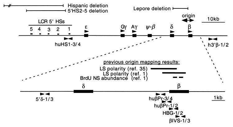
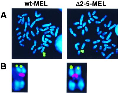
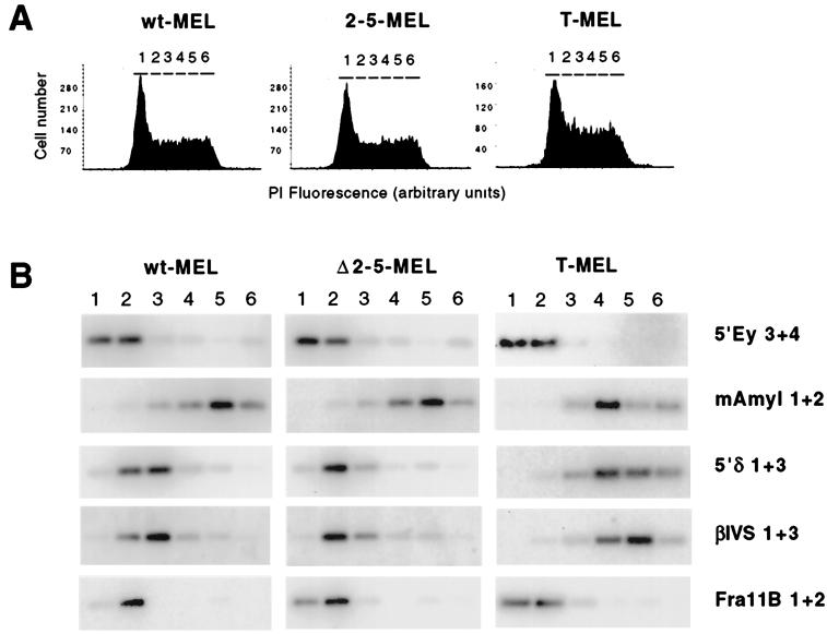
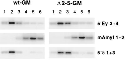
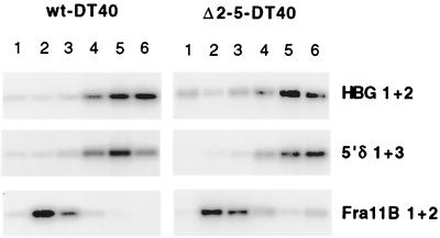
DNA replication in the human beta-globin locus is subject to long-distance regulation. In murine and human erythroid cells, the human locus replicates in early S phase from a bidirectional origin located near the beta-globin gene. This Hispanic thalassemia deletion removes regulatory sequences located over 52 kb from the origin, resulting in replication of the locus from a different origin, a shift in replication timing to late S phase, adoption of a closed chromatin conformation, and silencing of globin gene expression in murine erythroid cells. The sequences deleted include nuclease-hypersensitive sites 2 to 5 (5'HS2-5) of the locus control region (LCR) plus an additional 27-kb upstream region. We tested a targeted deletion of 5'HS2-5 in the normal chromosomal context of the human beta-globin locus to determine the role of these elements in replication origin choice and replication timing. We demonstrate that the 5'HS2-5-deleted locus initiates replication at the appropriate origin and with normal timing in murine erythroid cells, and therefore we conclude that 5'HS2-5 in the classically defined LCR do not control replication in the human beta-globin locus. Recent studies also show that targeted deletion of 5'HS2-5 results in a locus that lacks globin gene expression yet retains an open chromatin conformation. Thus, the replication timing of the locus is closely correlated with nuclease sensitivity but not globin gene expression.


Figures

FIG. 1
FIG. 1
Organization of the human β-globin locus. The positions of the globin gene reading frames and LCR DNase I-hypersensitive sites are indicated. The regions removed by the Hispanic, Lepore, and 5′HS2-5 deletions are denoted by lines. The bidirectional origin used to replicate the entire locus is located in the region of the β gene. Results of previous origin-mapping assays in the wild-type locus are summarized in the expanded view of the origin region; leading-strand polarity assays and a nascent strand abundance assay yield results in good agreement. The primer pairs used in this study to assay replication timing and origin activity are indicated by solid arrowheads.
FIG. 2
FIG. 2
Cytogenetic analysis of wt-MEL and Δ2-5-MEL hybrids. Metaphase chromosomes from each cell line were hybridized with (A) a human chromosome 11-specific paint (green) or (B) a human β-globin probe (green) in combination with a human centromeric probe (red). Chromosomes were counterstained with DAPI (4′,6′-diamidino-2-phenylindole) (blue). Both cell lines contain a single intact human chromosome 11 with the human β-globin locus in its normal position near the telomere of 11p.
FIG. 3
FIG. 3
Quantitative analysis of control duplex PCR products from the human β-globin locus in MEL hybrids. Amplification products from 2 ng of wt-MEL genomic DNA following 27, 28, 29, and 30 cycles of duplex PCR with primer pair 5′δ1+3 in combination with huβPr3+4 (A), huβPr1+2 (B), HBG1+2 (C), and βIVS1+3 (D) are shown; each reaction was performed in duplicate. Quantitation of these products reveals that in each case, the total amounts of product (bars) increase linearly with cycle number and the product ratios (lines) do not vary significantly between identical reactions or as the cycle number increases. These results demonstrate that each primer is in the linear phase of amplification.
FIG. 4
FIG. 4
DNA replication initiation in wild-type and mutant human β-globin loci in MEL hybrids. (A) Representative duplex PCR results from lambda exonuclease-resistant nascent strands prepared from wt-MEL and Δ2-5-MEL. Short nascent strands (ns1 to ns3; see Materials and Methods) and genomic DNA (G) from each cell line were used as templates for duplex PCR with one primer pair in the origin region in combination with a primer pair distant from the origin (5′δ1+3). Compared to genomic DNA, nascent strands from both cell lines are enriched for HBG1+2 and huβPr1+2 sequences, the primer pairs located centrally in the initiation region depicted in Fig. 1. Little or no enrichment is seen with flanking primer sets huβPr3+4 and βIVS1+3. (B) Quantification of duplex PCR analysis of wt-MEL and Δ2-5-MEL nascent strands. Three independent PCR analyses were performed; the mean value and standard error of the mean are plotted. A value above 1.0 represents enrichment of the origin-proximal sequences. The wild-type and Δ2-5 loci yield the same pattern: huβPr1+2 and HBG1+2 products are enriched, while products from adjacent primers show little or no enrichment. An independent nascent strand preparation from each hybrid yielded similar results. (C) Duplex PCR analysis of T-MEL (Hispanic deletion) nascent strands. Primers used are from the human (upper panel) and mouse (lower panel) β-globin loci. Primers in the human locus yield almost no products from nascent strands, despite strong amplification of genomic DNA with the same PCR premix; the signal strength from the nascent strands was not high enough above background to allow a ratio to be reliably determined. This PCR was repeated with the same result. In contrast, primers in the mouse locus yield comparable amounts of products from the nascent strands and genomic DNA, despite the fact that half the amount of nascent strand template was used for the mouse PCR as was used for the human PCR. Note the enrichment of 3′βmajor1+2 relative to LCR1+2 sequences, consistent with the location of the β-major primers within the initiation zone in the mouse locus (see text).
FIG. 5
FIG. 5
Replication timing of wild-type and mutant human β-globin loci in MEL hybrids. (A) Histogram of propidium iodide (PI) staining intensity (DNA content) of cells sorted for timing analysis. The gates used to sort cells into compartments, corresponding approximately to G1, S1 to S4, and G2, are labeled 1 to 6, respectively. (B) PCR and Southern analysis of replication timing in β-globin and control loci. Analysis was performed as described in the text, using primers for the human β-globin locus (5′δ1+3 and βIVS1+3), early-replicating control loci (endogenous murine β-globin [5′Ey3+4] and human cbl2 [Fra11B1+2]), and a late-replicating control locus (murine amylase, mAmyl1+2). In wt-MEL, the human β-globin locus replicates early (concurrently with murine β-globin and human Fra11B and earlier than amylase). In Δ2-5-MEL, the human β-globin locus replicates early, with a temporal profile similar to that seen in wt-MEL. Minor differences in timing between the human β-globin loci in wt-MEL and Δ2-5-MEL are also seen in the early controls and thus do not represent a difference in replication timing (see text). Analysis of a second independent Δ2-5-MEL clone yielded similar results. In contrast to wt-MEL and Δ2-5-MEL, the Hispanic deletion locus in T-MEL is late replicating.
FIG. 6
FIG. 6
Replication timing of wild-type and Δ2-5 loci in GM979 hybrids. Replication timing was assayed as in the MEL hybrids (see legend to Fig. 5). In both wt-GM and Δ2-5-GM, the human β-globin locus replicates early, relative to the early and late controls.
FIG. 7
FIG. 7
Replication timing of wild-type and Δ2-5 loci in DT40 hybrids. Replication timing assays were performed as in MEL and GM979 hybrids. In DT40, both the wild-type and Δ2-5 human β-globin loci (HBG1+2 and 5′δ1+3) are late replicating relative to the early-replicating Fra11B control.

References

    1. Aladjem M I, Groudine M, Brody L L, Dieken E S, Fournier R E, Wahl G M, Epner E M. Participation of the human beta-globin locus control region in initiation of DNA replication. Science. 1995;270:815–819. - PubMed
    1. Aladjem M I, Rodewald L W, Kolman J L, Wahl G M. Genetic dissection of a mammalian replicator in the human beta-globin locus. Science. 1998;281:1005–1009. - PubMed
    1. Bender M, Bulger M, Close J, Groudine M. Globin gene switching and DNaseI sensitivity of the endogenous β globin locus in mice do not require the locus control region. Mol Cell. 2000;5:387–393. - PubMed
    1. Bergemann A D, Johnson E M. The HeLa Pur factor binds single-stranded DNA at a specific element conserved in gene flanking regions and origins of DNA replication. Mol Cell Biol. 1992;12:1257–1265. - PMC - PubMed
    1. Brown B A, Padgett R W, Hardies S C, Hutchison C A, Edgell M H. Beta-globin transcript found in induced murine erythroleukemia cells is homologous to the beta h0 and beta h1 genes. Proc Natl Acad Sci USA. 1982;79:2753–2757. - PMC - PubMed

Publication types

LinkOut - more resources