Case Reports
. 2000 Aug;20(15):5653-64.
doi: 10.1128/MCB.20.15.5653-5664.2000.

Mutations in conserved regions of the predicted RAG2 kelch repeats block initiation of V(D)J recombination and result in primary immunodeficiencies

Affiliations
Case Reports

Mutations in conserved regions of the predicted RAG2 kelch repeats block initiation of V(D)J recombination and result in primary immunodeficiencies

C A Gomez et al. Mol Cell Biol. 2000 Aug.

Abstract

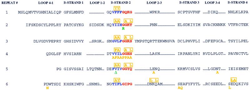
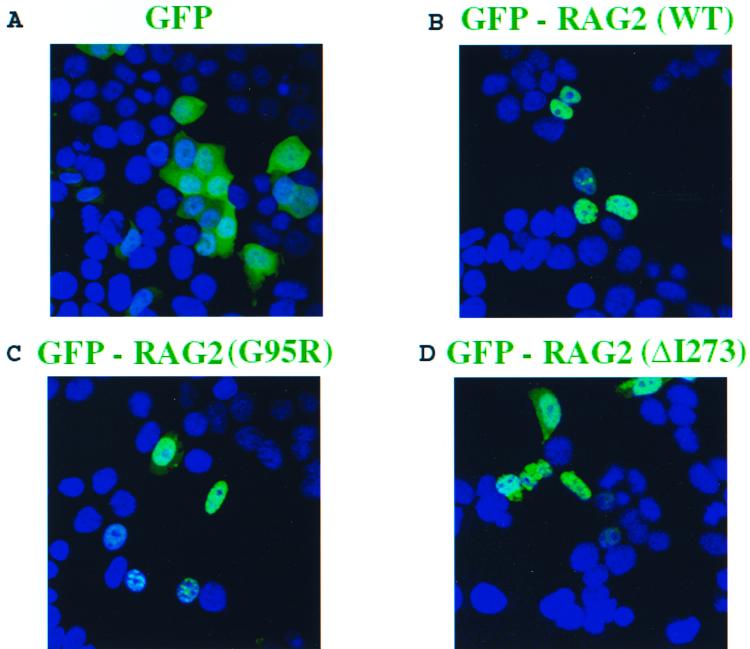
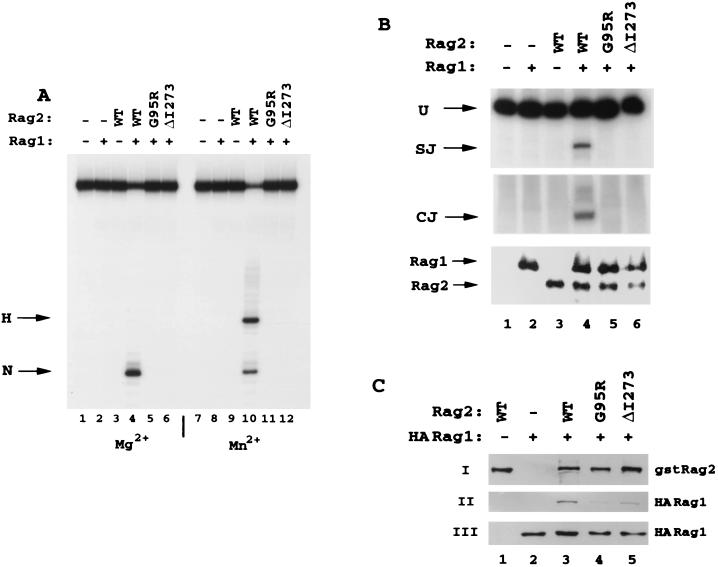
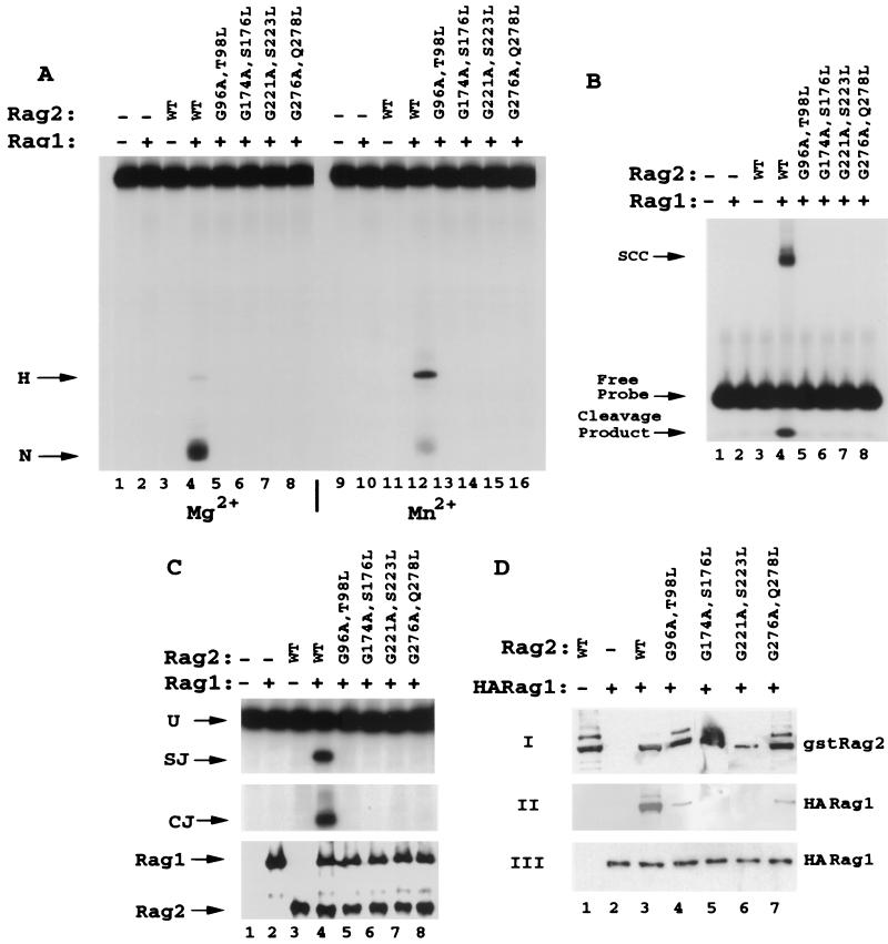
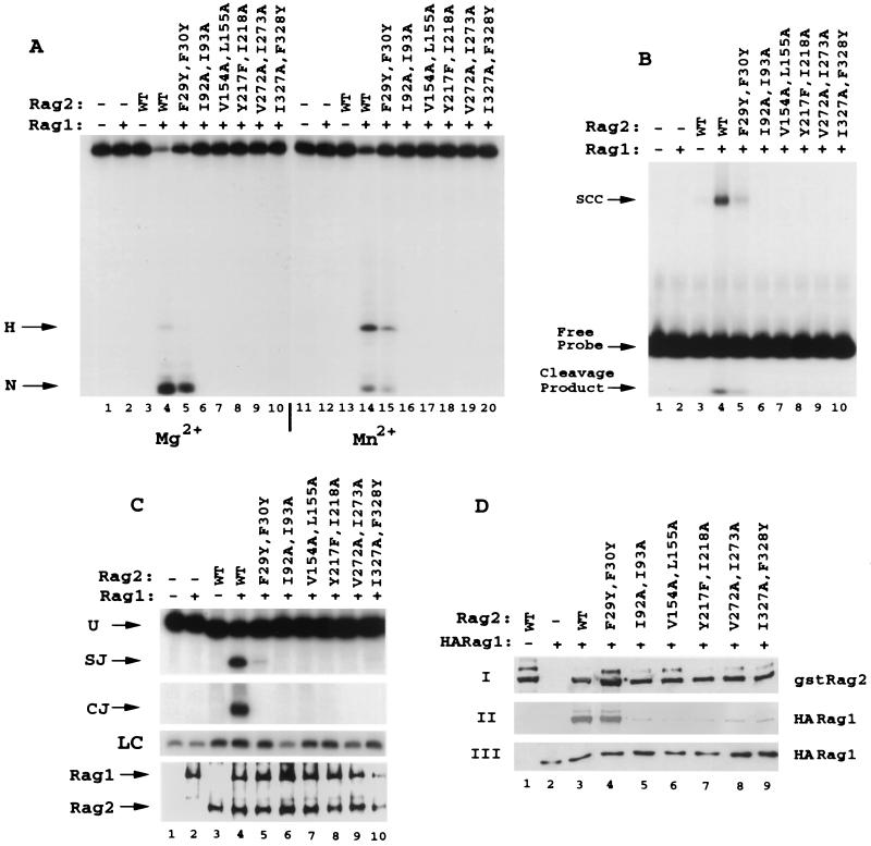
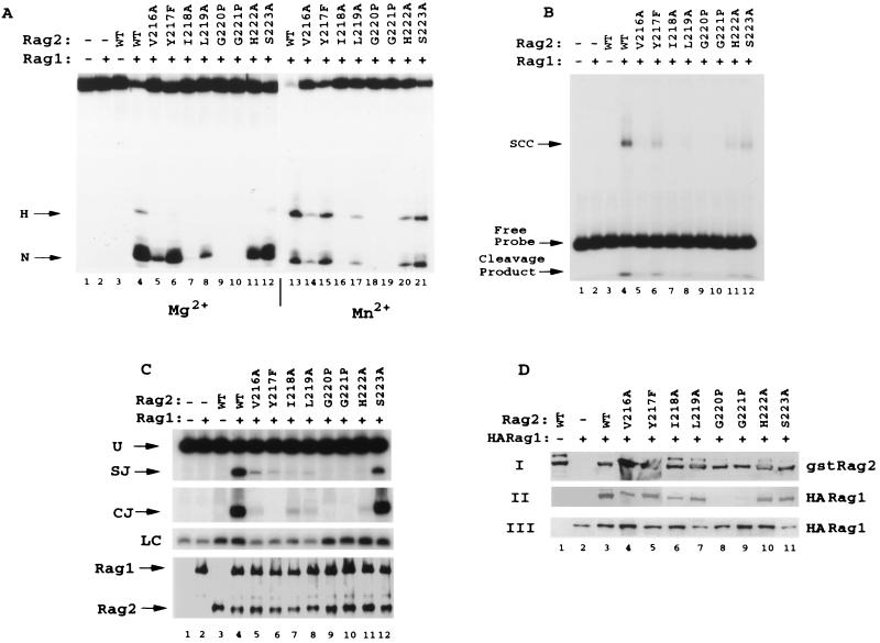
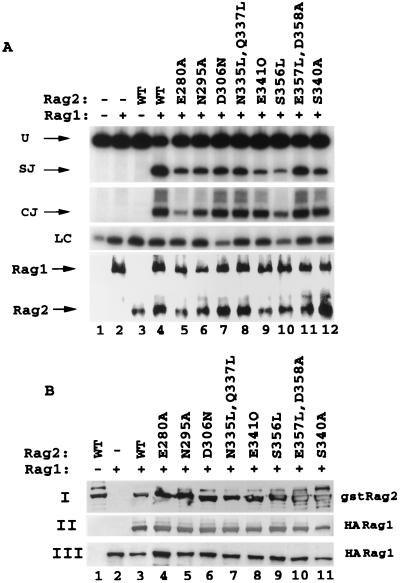
The V(D)J recombination reaction is composed of multiple nucleolytic processing steps mediated by the recombination-activating proteins RAG1 and RAG2. Sequence analysis has suggested that RAG2 contains six kelch repeat motifs that are predicted to form a six-bladed beta-propeller structure, with the second beta-strand of each repeat demonstrating marked conservation both within and between kelch repeat-containing proteins. Here we demonstrate that mutations G95R and DeltaI273 within the predicted second beta-strand of repeats 2 and 5 of RAG2 lead to immunodeficiency in patients P1 and P2. Green fluorescent protein fusions with the mutant proteins reveal appropriate localization to the nucleus. However, both mutations reduce the capacity of RAG2 to interact with RAG1 and block recombination signal cleavage, therefore implicating a defect in the early steps of the recombination reaction as the basis of the clinical phenotype. The present experiments, performed with an extensive panel of site-directed mutations within each of the six kelch motifs, further support the critical role of both hydrophobic and glycine-rich regions within the second beta-strand for RAG1-RAG2 interaction and recombination signal recognition and cleavage. In contrast, multiple mutations within the variable-loop regions of the kelch repeats had either mild or no effects on RAG1-RAG2 interaction and hence on the ability to mediate recombination. In all, the data demonstrate a critical role of the RAG2 kelch repeats for V(D)J recombination and highlight the importance of the conserved elements of the kelch motif.


Figures

FIG. 1
FIG. 1
Mutations in patients P1 and P2 are localized to the RAG2 kelch repeat domains. Each of the six kelch repeats of the RAG2 active core (13, 40, 41, 52) is formed by four β-strands (1 to 4) separated by loops of variable length (4-1 to 3-4). We present the sequence of mouse RAG2. The second β-strand of each repeat demonstrates the highest level of conservation between various members of the kelch family and is composed of a 4-amino-acid (predominantly hydrophobic) region, displayed in blue. The border of β-strand 2 and loop 2-3 contains a 4-amino-acid glycine-serine-threonine-rich repeat, highlighted in red. Mutations from patients P1 and P2 are noted in green below the affected residue. Note that isoleucine 273 in human RAG2 corresponds to a valine in the mouse sequence. Individual site-directed mutations are displayed in orange below the substituted amino acid, while multiple mutations are indicated and boxed in orange above the altered amino acids.
FIG. 2
FIG. 2
Mutations G95R and ΔI273 are imported into the nucleus. Various full-length RAG2 alleles (amino acids 1 to 527) were fused in frame to the C terminus of enhanced GFP and transiently overexpressed in 293T cells. The cells were fixed and processed 24 h posttransfection and subsequently visualized using confocal laser-scanning microscopy. Nuclei were stained with DAP1 (blue). (A) Cells were transfected with GFP alone. (B to D) Full-length forms of wild-type or mutant GFP-RAG2 localized effectively to the nucleus (GFP-RAG2 wild type [B], GFP-RAG2 G95R [C], and GFP-RAG2 ΔI273 [D]).
FIG. 3
FIG. 3
Human mutations in the predicted second β-strand of the kelch repeats are defective for both in vitro RSS cleavage and in vivo recombination activity. (A) Wild-type full-length human RAG2 and mutants G95R and ΔI273 were purified from 293T cells as fusion proteins to the C terminus of GST. Equal amounts of each protein were incubated with wild-type (WT) GST-RAG1 (amino acids 380 to 1040) and tested for the ability to generate nicks (N) and hairpins (H) on a 53-bp oligonucleotide containing a consensus 12 RSS. The DNA was 32P labeled on the 5′ end of the nicked strand. Reactions were conducted for 30 min at 37°C in the presence of Mg2+ (lanes 1 to 6) or Mn2+ (lanes 7 to 12), and the products were resolved on denaturing polyacrylamide gels. (B) Plasmids expressing the GST fusions of RAG2 and wild-type RAG1 were cotranfected into 293T cells along with a recombination plasmid substrate, pJH200. Recombination activity was evaluated by PCR analysis for the formation of signal joints (SJ, top panel) and coding joints (CJ, middle panel) of recombination substrate recovered 48 h posttransfection (39). PCR primers for signal joints also detect unrecombined plasmid (U). Dilutions of each sample were used to determine the linear range for the PCR analysis, and recombination activity was analyzed within this range. Protein levels were monitored by Western blot analysis of an aliquot of cell lysate (lower panel). (C) GST-RAG2 wild-type and mutant proteins were expressed with HA-tagged RAG1 active core (amino acids 330 to 1040), and interaction was evaluated by coprecipitation assays using glutathione beads. Panel I was blotted with anti-GST antibodies for detection of affinity-purified GST-RAG2, panel II was blotted with anti-HA antibody for detection of precipitated HA-RAG1, and panel III was blotted with polyclonal anti-RAG1 antibody R1P7.
FIG. 4
FIG. 4
Mutations in RAG2 glycine-serine-threonine-rich regions affect the interaction with RAG1 and concomitantly block RSS binding and nicking. (A) Double mutations in repeats 2 (G96A T98L), 3 (G174A S176L), 4 (G221A S223L), and 5 (G276A Q278L) were generated by substituting the second glycine of the glycine doublets as well as a serine or threonine residue 2 amino acids C-terminal to the glycine (except in the case of the fifth repeat, which has glutamine at this position [Fig. 1]). GST-RAG2 fusions (amino acids 1 to 383) were purified and tested for 12 RSS nicking and hairpin activity. In both Mg2+ (lanes 1 to 8) and Mn2+ (lanes 9 to 16), all four mutant proteins were entirely inactive for either nicking or hairpinning the 12 RSS compared to the wild type (WT). (B) Mutant RAG2 proteins were assayed for the capacity to form SCC along with wild-type GST-RAG1 on the 12 RSS. As with 12 RSS cleavage, all mutants were inactive for SCC formation (lanes 5 to 8). Cleavage products can be visualized below the free probe. (C) Mutants were tested for recombination activity on substrate pJH200 by PCR analysis for signal joints (SJ) and coding joints (CJ). None of the four mutants (lanes 5 to 8) generated detectable recombination products (SJ or CJ). Aliquots of the cell lysates were evaluated for protein expression by Western blot analysis. (D) Interaction between GST-RAG2 and HA-tagged RAG1 was monitored as described in the legend for Fig. 3C.
FIG. 5
FIG. 5
Mutation of most of the hydrophobic residues in the predicted second β-strand of repeats 1 to 6 of RAG2 decreases the binding of RAG1, with parallel effects on in vitro RSS binding and cleavage. (A and B) Conservative double mutations (F29Y F30Y, I92A I93A, V154A L155A, Y217F I218A, V272A I273A, and I327A F328Y) (Fig. 1) in the hydrophobic regions of kelch repeats 1 through 6 were expressed and purified as GST fusion proteins and tested in vitro for 12 RSS cleavage in Mg2+ (lanes 1 to 10) and Mn2+ (lanes 11 to 20) (A) or for SCC formation with the 12 RSS (B). (C and D) In vivo analysis of signal joint and coding-joint formation was conducted (C) in addition to analysis of complex formation between RAG1 and RAG2 (D). In panel C, loading controls (LC) performed in the linear range of the PCR are shown. WT, wild type.
FIG. 6
FIG. 6
Individual point mutations in the predicted second β-strand of repeat 4 have differential effects on interaction with RAG1 and on cleavage and recombination activity. Eight individual conservative point mutations (V216A, Y217F, I218A, L219A, G220P, G221P, H222A, and S223A) were produced in the second β-strand of the fourth kelch repeat of GST-RAG2 (amino acids 1 to 383). Mutants were analyzed for 12 RSS cleavage (A), binding (B), recombination activity (C), and RAG1 interaction (D). Panel C contains PCR loading controls (LC) performed in the linear range of the reaction. WT, wild type.
FIG. 7
FIG. 7
Mutations in predicted loop regions and at the predicted borders of β-strand 3 have moderate to no effect on the interaction with RAG1 and hence on the capacity to form coding and signal joints in vivo. Eight drastic mutations, E280A, N295A, D306N, N335L-Q337L, E341Q, S356L, E357L-D358A, and S340A, were introduced into the variable predicted loop regions of RAG2, and these substitutions were tested for the ability to mediate signal joint and coding-joint formation in vivo (A) and for the capacity to precipitate RAG1 (B). WT, wild type.

Similar articles

Cited by

References

    1. Adams J, Kelso R, Cooley L. The kelch repeat superfamily of proteins: propellers of cell function. Trends Cell Biol. 2000;10:17–24. - PubMed
    1. Agrawal A, Eastman Q M, Schatz D G. Transposition mediated by RAG1 and RAG2 and its implications for the evolution of the immune system. Nature. 1998;394:744–751. - PubMed
    1. Aidinis V, Bonaldi T, Beltrame M, Santagata S, Bianchi M E, Spanopoulou E. The RAG1 homeodomain recruits HMG1 and HMG2 to facilitate recombination signal sequence binding and to enhance the intrinsic DNA-bending activity of RAG1-RAG2. Mol Cell Biol. 1999;19:6532–6542. - PMC - PubMed
    1. Aidinis V, Dias D C, Gomez C A, Bhattacharyya D, Spanopoulou E, Santagata S. Definition of minimal domains of interaction within the recombination-activating genes 1 and 2 recombinase complex. J Immunol. 2000;164:5826–5832. - PubMed
    1. Akamatsu Y, Oettinger M A. Distinct roles of RAG1 and RAG2 in binding the V(D)J recombination signal sequences. Mol Cell Biol. 1998;18:4670–4678. - PMC - PubMed

Publication types

LinkOut - more resources