

Review
doi: 10.1083/jcb.150.1.f13.
AAA proteins. Lords of the ring
Affiliations
- PMID: 10893253
- PMCID: PMC2185557
- DOI: 10.1083/jcb.150.1.f13
Item in Clipboard
Review
AAA proteins. Lords of the ring
J Cell Biol.
.
No abstract available
Figures
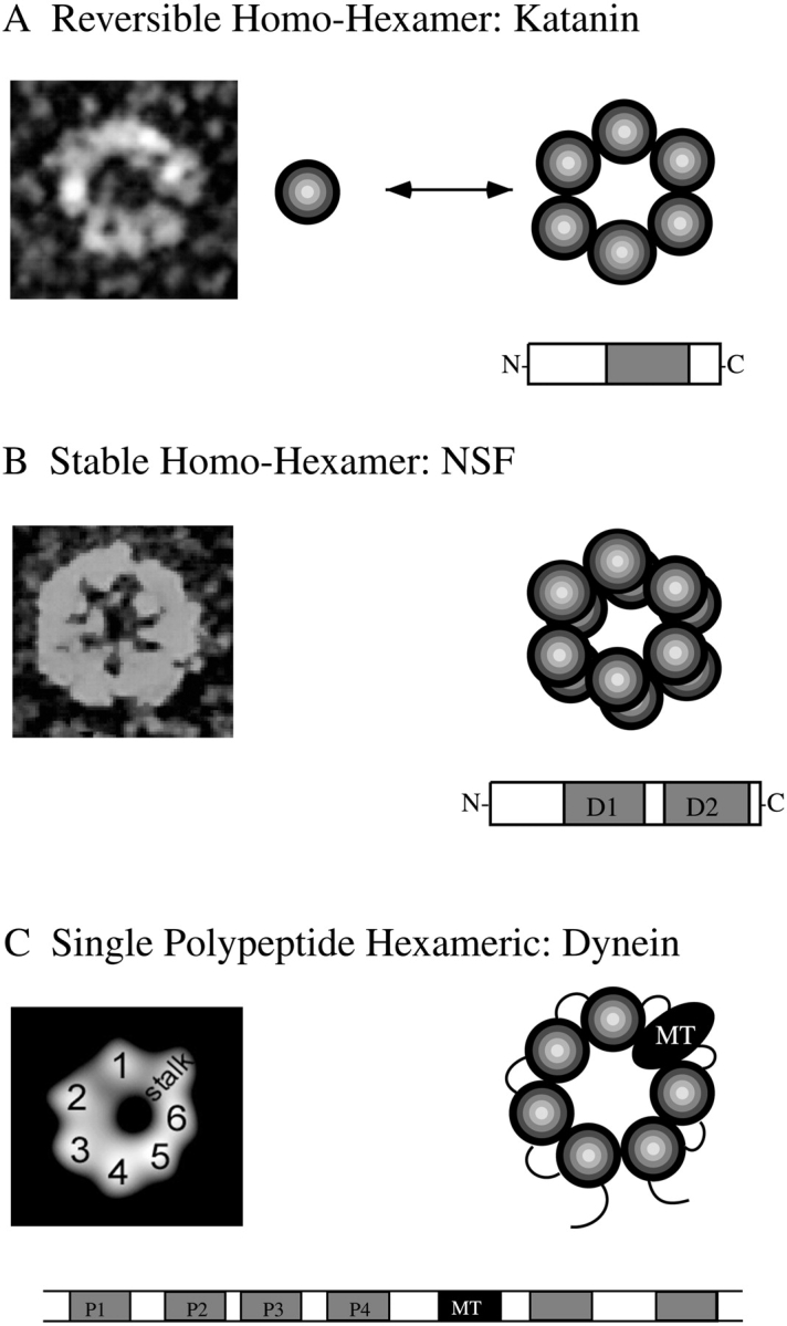
Three strategies for creating rings out of AAA domains. (A) Some single domain AAA proteins (katanin, VPS4) exist in an equilibrium between monomers and rings. Bound ATP (or nonhydrolyzable ATP analogues) as well as target protein binding favor ring formation. The NH2-terminal domain (not shown on the ring structure) is involved in target protein binding. (B) A second class of AAA proteins (such as NSF) have two AAA domains, one of which serves as the main hydrolytic domain and a second which binds ATP, but hydrolyzes it very poorly, and is involved in creating a stable hexamer base. The NH2-terminal domain of NSF also is involved in target protein (αSNAP) binding. (C) A third type of AAA protein (dynein) has six AAA domains within a single polypeptide chain as well as an additional microtubule binding domain. Four of these sites (P1-P4) bind ATP, and P1 acts as the main hydrolytic site. Electron micrographs of katanin and NSF were provided by J. Heuser, and the two-dimensional projection of the dynein motor domain (from single particle averaging of electron microscopic images) was provided by M. Samso and M. Koonce. The diameter of these AAA hexameric rings is ∼15 nm.
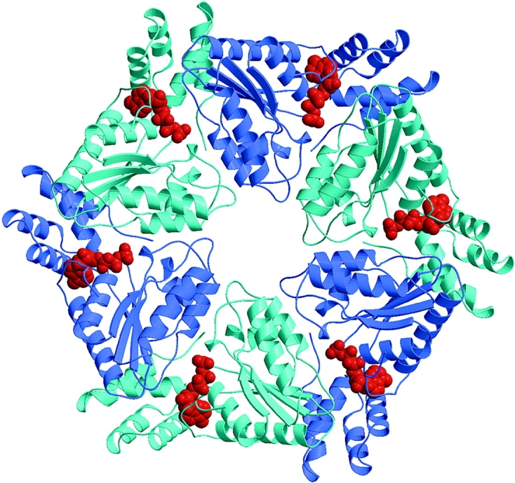
Atomic structure of the D2 domain of NSF in a hexameric ring. All subunits are identical, but adjacent subunits are colored in different shades of blue to illustrate the subunits interface. The nucleotide (AMPPNP shown as a red space-filling model) is located close to the interface between subunits, and subunit–subunit interactions are likely to affect the ATPase cycle. Atomic coordinates were provided by W. Weis (Stanford University).
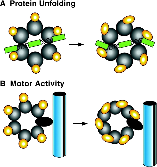
Speculative models for conformational changes of AAA proteins. (A) Target binding domains undergo a concerted, twisting conformational change. If a protein or protein complex is bound at two or more sites, it will be subject to mechanical stain by the conformational change. (B) A model for dynein-based motility based upon a conformational change in the ring. In this case, the shift in the AAA ring alters the angle of an embedded microtubule binding domain, which in turn produces a force that moves a microtubule. The dark gray spheres are the AAA domains and the smaller yellow sphere represent elements outside of the AAA domains (relative sizes are not necessarily accurate). Although not shown here, a conformational change may also have a vectoral component directed towards the center of the ring, as discussed in the text.
References
-
- Arlt H., Tauer R., Feldmann H., Neupert W., Langer T. The YTA10-12 complex, an AAA protease with chaperone-like activity in the inner membrane of mitochondria. Cell. 1996;85:875–885. - PubMed
-
- Baumeister W., Walz J., Zuhl F., Seemuller E. The proteasomeparadigm of a self-compartmentalizing protease. Cell. 1998;92:367–380. - PubMed
Publication types
MeSH terms
Substances
Grants and funding
LinkOut - more resources
Full Text Sources
Other Literature Sources
Molecular Biology Databases
