. 2000 Jul 10;150(1):77-88.
doi: 10.1083/jcb.150.1.77.

The unfolded protein response regulates multiple aspects of secretory and membrane protein biogenesis and endoplasmic reticulum quality control

Affiliations

The unfolded protein response regulates multiple aspects of secretory and membrane protein biogenesis and endoplasmic reticulum quality control

D T Ng et al. J Cell Biol. .

Abstract

The unfolded protein response (UPR) is an intracellular signaling pathway that relays signals from the lumen of the ER to activate target genes in the nucleus. We devised a genetic screen in the yeast Saccharomyces cerevisiae to isolate mutants that are dependent on activation of the pathway for viability. Using this strategy, we isolated mutants affecting various aspects of ER function, including protein translocation, folding, glycosylation, glycosylphosphatidylinositol modification, and ER-associated protein degradation (ERAD). Extending results gleaned from the genetic studies, we demonstrate that the UPR regulates trafficking of proteins at the translocon to balance the needs of biosynthesis and ERAD. The approach also revealed connections of the UPR to other regulatory pathways. In particular, we identified SON1/RPN4, a recently described transcriptional regulator for genes encoding subunits of the proteasome. Our genetic strategy, therefore, offers a powerful means to provide insight into the physiology of the UPR and to identify novel genes with roles in many aspects of secretory and membrane protein biogenesis.


Figures
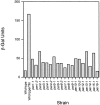
Figure 1
Figure 1
per mutants activate the unfolded protein response pathway. Wild-type and mutant cells carrying the UPRE-LacZ reporter gene are grown logarithmically and lysed. β-galactosidase activity is measured from lysates using 2-nitrophenyl-β-d-galactopyranoside (ONPG) as substrate and detected as an increase in absorbance at 420 nM. TM, cells treated with 2.5 μg/ml tunicamycin for 60 min before lysis.
Figure 3
Figure 3
A, per5-1 is defective in N-linked glycosylation. Wild-type (W303a) and per5-1 cells were metabolically pulse-labeled for 5 min and chased for 0, 15, and 30 min. CPY was immunoprecipitated from detergent lysates and divided into two aliquots for each time point. One aliquot was treated with endoglycosidase H and the other was mock-treated. Proteins were separated on a 10% polyacrylamide gel and visualized by fluorography. The positions of the different pro and mature glycoforms of CPY are indicated as described in Fig. 2. Endo H deglycosylated pro (dg-proCPY) and mature forms (dg-mCPY) are indicated to the right. B, per5-1 is an allele of RFT1. Wild-type, per5-1, and per5-1 (pDN387) cells were pulse-labeled for 5 min and chased for 0 and 30 min. pDN387 is a centromeric vector containing the RFT1 gene. Immunoprecipitates of Gas1p were resolved by a 10–15% polyacrylamide gradient gel and visualized by autoradiography. The normal ER and mature (Golgi/PM) forms are shown.
Figure 2
Figure 2
Processing of Gas1p and CPY in per mutants. Logarithmically growing wild-type and mutant cells were metabolically labeled with [35S]methionine/cysteine for 5 min at 30°C, followed by a chase for 30 min. From detergent lysates, Gas1p and CPY were immunoprecipitated using monospecific polyclonal antisera. As a control, these proteins were also immunoprecipitated from wild-type lysates pulse-labeled with no chase to indicate the ER forms (lane 1). Proteins are separated on a 10–15% polyacrylamide gradient gel and visualized by fluorography. The positions of preGas1p, ER Gas1p, and Golgi/plasma membrane Gas1p (Golgi/PM) are indicated (A). For CPY, the ER (P1), Golgi apparatus (P2), and mature vacuolar form (CPY) are indicated to the left and the vacuolar underglycosylated forms (-1, -2, -3, -4) to the right of B.
Figure 4
Figure 4
PER5/RFT1 is a UPR target gene. A, Alignment of UPRE sequences. Experimentally determined UPRE sequences (Mori et al. 1998) are shown with an upstream sequence of PER5/RFT1 that conforms to the palindromic motif shown by the arrows. Dots denote additional important positions in the motif. B, Northern analysis using RNA extracted from W303a, DNY421, and Δhac1 cells incubated in the presence or absence of 2.5 μg/ml of tunicamycin for 60 min at 30°C. PER5/RFT1, KAR2, and ACT1 transcripts were blotted onto a nylon filter, hybridized to 32P-labeled probes sequentially, and visualized by autoradiography. Quantification was performed by PhosphorImager analysis.
Figure 5
Figure 5
A subset of per mutants is defective for ERAD. All twenty per mutants were transformed with pDN436 (CPY*HA) and pulse-chase analysis performed to measure the degradation of the ERAD substrate CPY*HA. Immunoprecipitates of CPY*HA were normalized using TCA precipitable counts. The proteins were separated by PAGE followed by autoradiography. Quantification was performed by PhosphorImager analysis and reported as percent remaining to the right of each respective autoradiogram. In addition to the wild-type (DNY563) and ERAD mutant (DNY572, cue1::TRP1) controls, only per mutants with significant ERAD defects are shown.
Figure 6
Figure 6
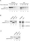
The UPR is required for efficient ER protein translocation and ERAD. A, Wild-type and ire1::TRP1 cells (labeled Δire1) expressing CPY*HA were pulse-labeled for 10 min with [35S]methionine/cysteine and chased for 0, 30, 60, and 90 min. CPY*HA was immunoprecipitated using anti-HA mAb and analyzed by SDS-PAGE. The proteins were visualized by direct autoradiography and quantification of the gel was performed using a PhosphorImager. B, Wild-type, sec62-101, and Δire1 cells expressing CPY*HA were pulse-labeled for 10 min and CPY*HA immunoprecipitated and separated on a 10% polyacrylamide gel (lanes 1, 2, and 3). Lanes 4 and 5, Gas1p immunoprecipitated from the lysates used for lanes 1 and 3. C, Wild-type, sec62-101, and Δire1cells expressing CPYHA were pulse-labeled for 10 min and immunoprecipitated for endogenous CPY. The position of preproCPYHA, the ER P1, and Golgi P2 forms are indicated.
Figure 7
Figure 7
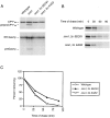
Increased gene dosage of specific UPR targets alleviate translocation and ERAD defects in UPR-deficient cells. A, Wild-type and Δire1 cells expressing CPY*HA were pulse-labeled with [35S]methionine/cysteine followed by immunoprecipitation for CPY*HA (top) and Gas1p (bottom). Haploid strains DNY563 (wild-type), ESY157 (Δire1), ESY158 (ESY157 with an extra copy of KAR2 on the centromeric plasmid pMR713; labeled Δire1, 2n KAR2), and ESY159 (ESY157 with and extra copy of SEC61 on the centromeric plasmid pCS15; labeled Δire1, 2n SEC61) were analyzed. The lumenal and nontranslocated forms (pre) are indicated. B, DNY563 (wild-type), ESY158 (Δire1, 2n KAR2), and ESY159 (Δire1, 2n SEC61) cells were pulse-labeled with [35S]methionine/cysteine for 10 min, followed by a cold chase for the times indicated, CPY*HA immunoprecipitated from detergent lysates, and separated by SDS-PAGE. C, Quantification of immunoprecipitated CPY*HA shown in B by PhosphorImager analysis.

References

    1. Baxter B.K., James P., Evans T., Craig E.A. SSI1 encodes a novel Hsp70 of the Saccharomyces cerevisiae endoplasmic reticulum. Mol. Cell. Biol. 1996;16:6444–6456. - PMC - PubMed
    1. Biederer T., Volkwein C., Sommer T. Degradation of subunits of the Sec61p complex, an integral component of the ER membrane, by the ubiquitin–proteasome pathway. EMBO (Eur. Mol. Biol. Organ.) J. 1996;15:2069–2076. - PMC - PubMed
    1. Biederer T., Volkwein C., Sommer T. Role of Cue1p in ubiquitination and degradation at the ER surface. Science. 1997;278:1806–1809. - PubMed
    1. Boeke J.D., LaCroute F., Fink G.R. A positive selection for mutants lacking orotidine-5′-phosphate decarboxylase activity in yeast5-fluoro-orotic acid resistance. Mol. Gen. Genet. 1984;197:345–346. - PubMed
    1. Brodsky J.L., McCracken A.A. ER protein quality control and proteasome-mediated protein degradation. Sem. Cell Dev. Biol. 1999;10:501–513. - PubMed

Publication types

MeSH terms