. 2000 Jul 10;150(1):89-103.
doi: 10.1083/jcb.150.1.89.

Molecular basis for Rab prenylation

Affiliations

Molecular basis for Rab prenylation

C Alory et al. J Cell Biol. .

Abstract

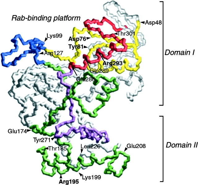
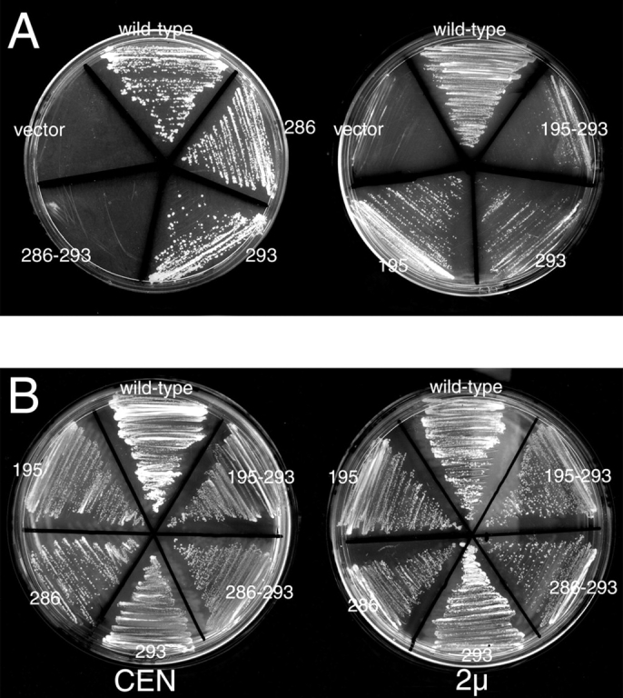
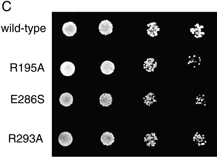
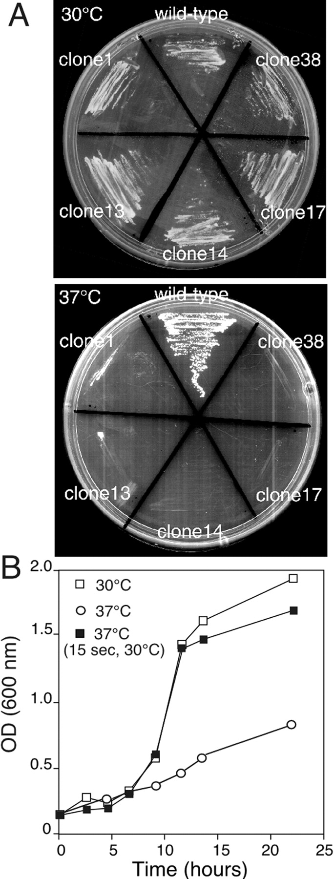
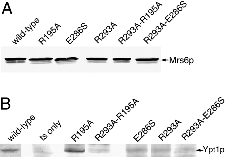
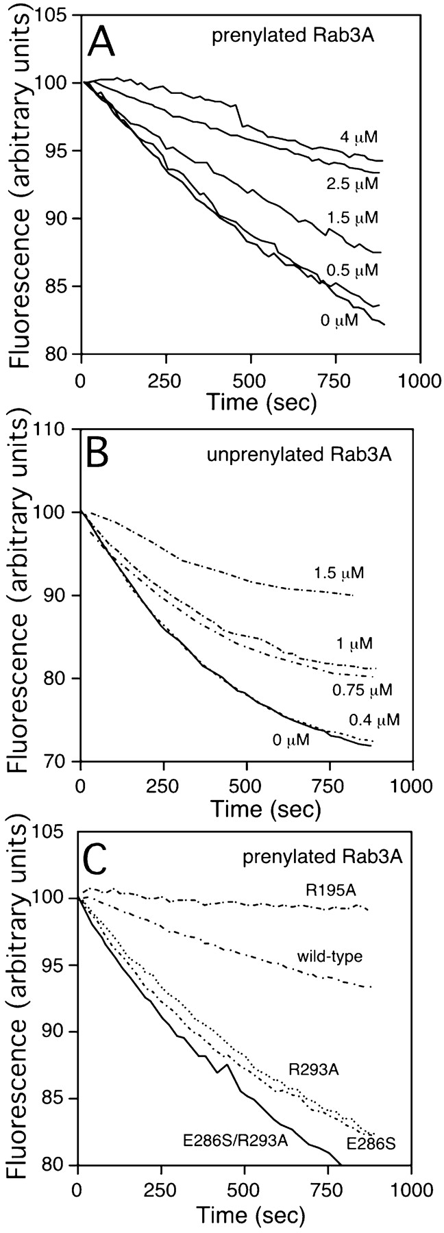
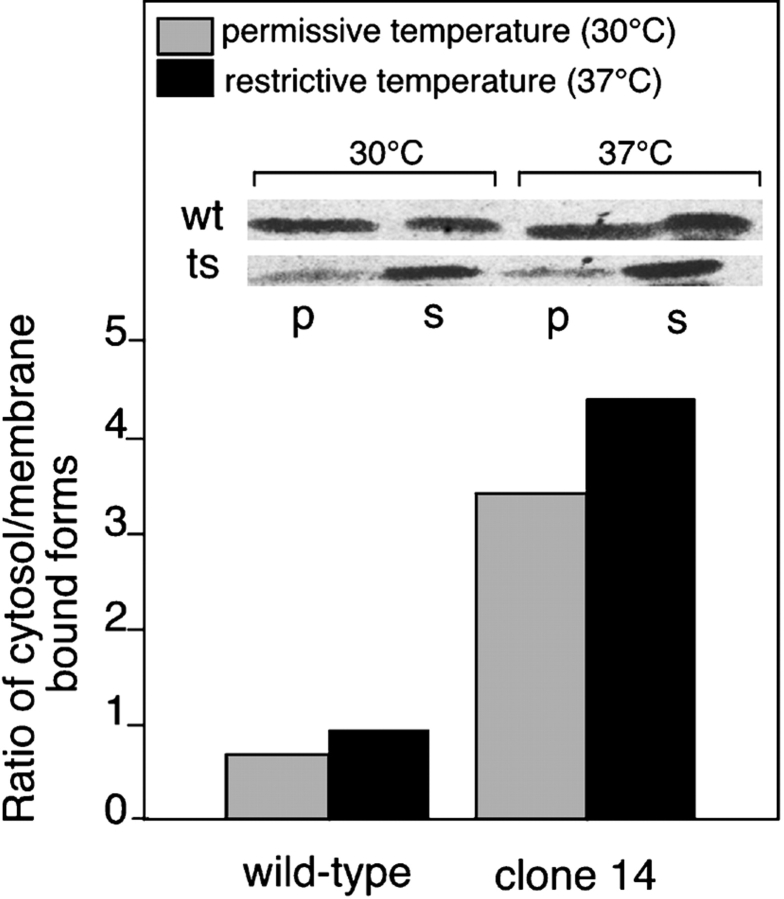
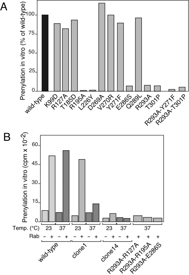
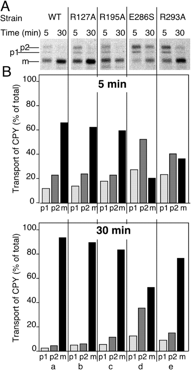
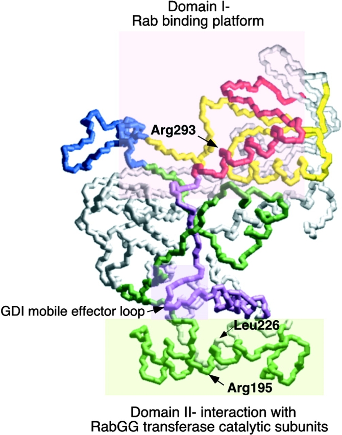
Rab escort proteins (REP) 1 and 2 are closely related mammalian proteins required for prenylation of newly synthesized Rab GTPases by the cytosolic heterodimeric Rab geranylgeranyl transferase II complex (RabGG transferase). REP1 in mammalian cells is the product of the choroideremia gene (CHM). CHM/REP1 deficiency in inherited disease leads to degeneration of retinal pigmented epithelium and loss of vision. We now show that amino acid residues required for Rab recognition are critical for function of the yeast REP homologue Mrs6p, an essential protein that shows 50% homology to mammalian REPs. Mutant Mrs6p unable to bind Rabs failed to complement growth of a mrs6Delta null strain and were found to be dominant inhibitors of growth in a wild-type MRS6 strain. Mutants were identified that did not affect Rab binding, yet prevented prenylation in vitro and failed to support growth of the mrs6Delta null strain. These results suggest that in the absence of Rab binding, REP interaction with RabGG transferase is maintained through Rab-independent binding sites, providing a molecular explanation for the kinetic properties of Rab prenylation in vitro. Analysis of the effects of thermoreversible temperature-sensitive (mrs6(ts)) mutants on vesicular traffic in vivo showed prenylation activity is only transiently required to maintain normal growth, a result promising for therapeutic approaches to disease.


Figures

Figure 1
Figure 1
Potential location of substitutions (yeast numbering) based on the crystal structure of bovine α-GDI. The tentative location of each of the residues (Mrs6p residue numbering) mutated in this study based on their homologous residue in α-GDI (Table ) are indicated (Schalk et al. 1996). The insert region (blue) and the Rab-binding region containing SCRs 1B (yellow) and 3B (red) are highlighted at the top of GDI and form domain I. The conserved face of GDI containing SCRs 2 (green) and 3A (purple) link domain I with a second domain II, and are oriented to the front of the image (Schalk et al. 1996). Bolded residues are those that have prominent phenotype in altering REP function.
Figure 2
Figure 2
Ability of selected single and double point mutants to complement growth of mrs6Δ null strain. A, Indicated mutants were expressed in the mrs6Δ null strain using the single copy CEN plasmid as described in Materials and Methods. B, Indicated mutants were expressed using the single copy CEN plasmid, or multicopy 2μ plasmid in wild-type cells. The ability of overexpressed Mrs6p double mutants to partially inhibit growth is illustrated by reduced level of colonies appearing on the plate. C, Indicated mutants were expressed in the mrs6Δ null strain using the single copy CEN plasmid as described in Materials and Methods. 5,000, 2,000, 500, and 50 cells were spotted onto a plate. The growth was assessed after a 2–4-d incubation at 30°C.
Figure 2
Figure 2
Ability of selected single and double point mutants to complement growth of mrs6Δ null strain. A, Indicated mutants were expressed in the mrs6Δ null strain using the single copy CEN plasmid as described in Materials and Methods. B, Indicated mutants were expressed using the single copy CEN plasmid, or multicopy 2μ plasmid in wild-type cells. The ability of overexpressed Mrs6p double mutants to partially inhibit growth is illustrated by reduced level of colonies appearing on the plate. C, Indicated mutants were expressed in the mrs6Δ null strain using the single copy CEN plasmid as described in Materials and Methods. 5,000, 2,000, 500, and 50 cells were spotted onto a plate. The growth was assessed after a 2–4-d incubation at 30°C.
Figure 3
Figure 3
Growth of the mrs6 ts strain. A, The indicated temperature-sensitive strains (clones 1, 13, 14, 17, and 38) were incubated at the permissive (30°C; left) or the restrictive (37°C; right) temperature. B, Growth of clone 14 at the permissive (open squares) or restrictive (open circles) temperatures. In the curve shown by the closed squares, the culture was exposed to the permissive temperature for 15 s before reequilibration to the restrictive temperature (∼2 min total time at the permissive temperature).
Figure 4
Figure 4
Coimmunoprecipitation of Ypt1p and Sec4p with wild-type and mutant HA-Mrs6p proteins. A and B, Identical amounts of cell homogenates of mrs6 ts cells (grown at 30°C [not shown] or 37°C) expressing HA epitope-tagged wild-type or mutant Mrs6p proteins (indicated at the top of each lane) were cleared of membranes by centrifugation at 100,000 g, and HA-Mrs6p was immunoprecipitated from each supernatant fraction (S100) under native conditions. In A, HA-Mrs6p was detected using immunoblotting with an HA-specific antibody in cell homogenates (before immunoprecipitation) to show that identical amounts of HA-Mrs6p are present in each sample. In B, antibodies to Ypt1p were used to detect Ypt1p in the immunoprecipitates using immunoblotting. All blots were treated in an identical fashion to allow quantitative comparison of levels of recovered Ypt1p.
Figure 5
Figure 5
Fluorescence assay to measure Mrs6p-Rab3A interactions in vitro. A and B, Effect of Mrs6p wild-type and mutants on the intrinsic dissociation of mant-GDP from Rab3A. Mrs6p was added at the indicated final concentration (μM). The interaction of Mrs6p with prenylated (A) and unprenylated (B) Rab3A was determined by measuring the decrease in relative fluorescence (λ excitation 360 nm, λ emission 440 nm) that accompanies the release of mant-GDP from Rab3A as described in Materials and Methods. C, The interaction of Mrs6p mutants with Rab3A was determined by mixing 100 nM Rab3A with 2.5 μM Mrs6p wild-type or mutant protein as indicated.
Figure 6
Figure 6
Distribution of wild-type and mutant Rab proteins between cytosol and membranes. Wild-type and clone 14 mrs6 ts strain were incubated at the indicated temperature. After 5 h, the same OD of cells was lysed and subjected to centrifugation at 100,000 g to generate a P100 membrane fraction (p) and an S100 cytosol fraction (s). The amount of Ypt1p in each fraction was quantitated by immunoblotting with specific antibody. The inset illustrates that the amount of total Rab at the 5-h time point that was detected using immunoblotting was the same in both the wild-type and ts strains at either 30°C or 37°C.
Figure 7
Figure 7
Analysis of prenylation activity of Mrs6p single and double mutants in vitro. A, Single and double mutants that partially or fully complemented growth were expressed in the mrs6Δ strain and extracts prepared for assay in the presence or absence of recombinant Ypt1p as described in Materials and Materials. B, To assess the prenylation activity of inviable double mutants, these were expressed in the mrs6 ts strain and extracts prepared for assay as indicated. The assay profile of clone 14 is typical for that observed for clones 13, 17, and 38. Double mutants were assayed in the clone 14 strain at 37°C.
Figure 8
Figure 8
Analysis of vesicle-mediated trafficking of mrs6 single mutants. The indicated strains in mrs6Δ null cells were prepared and the cultures were labeled with [35S]methionine/cysteine for 20 min, then chased for 5 or 30 min. CPY was immunoprecipitated from extracts derived from each culture. The positions of p1-CPY (ER), p2-CPY (Golgi), and mature (m) CPY (vacuole) are indicated to the left. In wild-type cells incubated at 37°C transport is very rapid. Here, CPY is largely recovered in the mature form, even after a 5-min chase period. B, The distribution of CPY in the p1, p2, and m forms at the 5-min (top) and 30-min (bottom) time points for each mutant were quantitated using densitometry.
Figure 9
Figure 9
EM of a mrs6 ts mutant strain. The clone 14 mrs6 ts mutant was grown overnight at 30°C, the culture was divided, and one half was incubated at 37°C for 3 h (C) while the other half was maintained at 30°C (B). In C, note the numerous large vesicles that accumulate (arrowheads), and the punched-in appearance of vacuoles (arrows) as a consequence of inactivation of Mrs6p. A typical wild-type cell is shown in A.
Figure 10
Figure 10
Domain organization of REP for Rab-binding and prenylation. The figure highlights principle regions involved in REP function based on the homologous residues in GDI through sequence alignment. Residues shaded by the pink box in upper region (Domain I) directs Rab interaction; residues shaded by the light green box in the lower region (Domain II) participate in recognition of the catalytic subunits of GG Tr II. The mobile effector loop involved in recognition of recycling factors by GDI (Luan et al. 2000), but not REP (this study), are indicated by the shaded purple box at the interface of domains I and II.

Similar articles

Cited by

References

    1. Alexandrov K., Horiuchi H., Steele-Mortimer O., Seabra M.C., Zerial M. Rab escort protein-1 is a multifunctional protein that accompanies newly prenylated rab proteins to their target membranes. EMBO (Eur. Mol. Biol. Organ.) J. 1994;13:5262–5273. - PMC - PubMed
    1. Alexandrov K., Simon I., Yurchenko V., Iakovenko A., Rostkova E., Scheidig A.J., Goody R.S. Characterization of the ternary complex between Rab7, REP-1 and Rab geranylgeranyl transferase. Eur. J. Biochem. 1999;265:160–170. - PubMed
    1. Anant J.S., Desnoyers L., Machius M., Demeler B., Hansen J.C., Westover K.D., Deisenhofer J., Seabra M.C. Mechanism of Rab geranylgeranylationformation of the catalytic ternary complex. Biochemistry. 1998;37:12559–12568. - PubMed
    1. Andres D.A., Seabra M.C., Brown M.S., Armstrong S.A., Smeland T.E., Cremers F.P.M., Goldstein J.L. cDNA cloning of component A of rab geranylgeranyl transferase and demonstration of its role as a Rab escort protein. Cell. 1993;73:1091–1099. - PubMed
    1. Cadwell R.C., Joyce G.F. Randomization of genes by PCR mutagenesis. PCR Methods Appl. 1992;2:28–33. - PubMed

Publication types

MeSH terms

Substances