. 2000 Aug;182(15):4268-77.
doi: 10.1128/JB.182.15.4268-4277.2000.

A redox-responsive regulator of photosynthesis gene expression in the cyanobacterium Synechocystis sp. Strain PCC 6803

Affiliations

A redox-responsive regulator of photosynthesis gene expression in the cyanobacterium Synechocystis sp. Strain PCC 6803

H Li et al. J Bacteriol. 2000 Aug.

Abstract

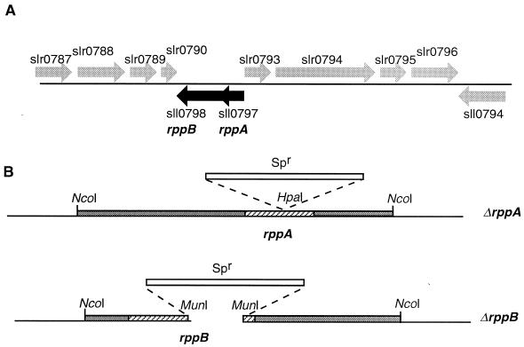
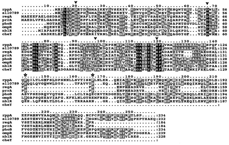
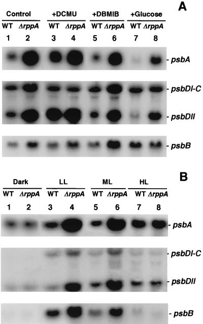
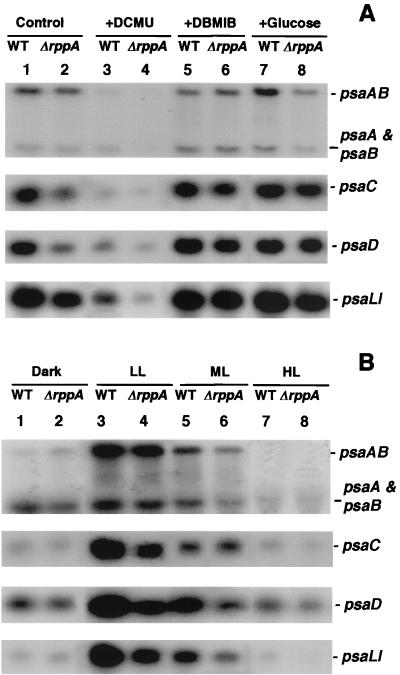
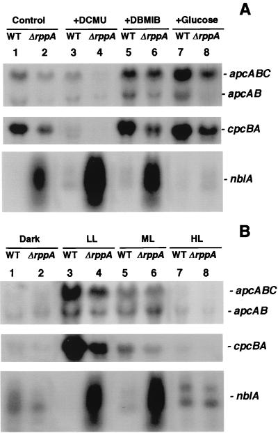
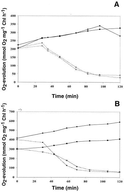
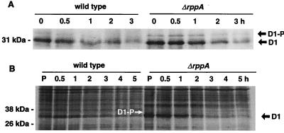
We have identified genes in the unicellular cyanobacterium Synechocystis sp. strain PCC 6803 that are involved with redox control of photosynthesis and pigment-related genes. The genes, rppA (sll0797) and rppB (sll0798), represent a two-component regulatory system that controls the synthesis of photosystem II (PSII) and PSI genes, in addition to photopigment-related genes. rppA (regulator of photosynthesis- and photopigment-related gene expression) and rppB exhibit strong sequence similarity to prokaryotic response regulators and histidine kinases, respectively. In the wild type, the steady-state mRNA levels of PSII reaction center genes increased when the plastoquinone (PQ) pool was oxidized and decreased when the PQ pool was reduced, whereas transcription of the PSI reaction center genes was affected in an opposite fashion. Such results suggested that the redox poise of the PQ pool is critical for regulation of the photosystem reaction center genes. In Delta rppA, an insertion mutation of rppA, the PSII gene transcripts were highly up-regulated relative to the wild type under all redox conditions, whereas transcription of phycobilisome-related genes and PSI genes was decreased. The higher transcription of the psbA gene in Delta rppA was manifest by higher translation of the D1 protein and a concomitant increase in O(2) evolution. The results demonstrated that RppA is a regulator of photosynthesis- and photopigment-related gene expression, is involved in the establishment of the appropriate stoichiometry between the photosystems, and can sense changes in the PQ redox poise.


Figures

FIG. 1
FIG. 1
Structure of the rpp region and mutations of rppA and rppB: genes of Synechocystis sp. strain PCC 6803. (A) Genomic organization of the response regulator gene rppA (locus sll0797) and the adjacent sensor kinase gene rppB (locus s110798). (B) Genetic construction of ΔrppA and ΔrppB. The rppA gene was interrupted by insertion of a spectinomycin resistance (Spr) cassette in the HpaI site; the rppB gene was inactivated by replacement of a 789-bp MunI-MunI fragment with the spectinomycin resistance cassette.
FIG. 2
FIG. 2
Alignment of RppA with other response regulators. The sequences are as follows: RppA and sll0789, Synechocystis sp. strain PCC 6803; RegA, Rhodobacter capsulatus (GenBank accession no. M64976); PrrA, R. sphaeroides (L25895); PhoB, Synechococcus sp. WH7803 (U38917); OmpR, Escherichia coli (J01656); NblR, Synechococcus sp. strain PCC 7942 (AF049128); CheY, E. coli (M13463). Identical residues are shaded in black, conserved residues are shaded in dark gray, and similar residues are shaded in light gray. Arrowheads denote the highly conserved aspartic acid (Asp8 and Asp53), threonine (Thr81), and lysine (Lys103) residues in RppA, which correspond to Asp13, Asp57, Thr87, and Lys109 in CheY, as well as to Asp20, Asp63, Thr91, and Lys113 in PrrA/RegA. Stars denote the two conserved prolines in RppA, s110789, RegA, and PrrA.
FIG. 3
FIG. 3
Northern blot analysis of PSII gene expression in Synechocystis sp. strain PCC 6803 wild type (WT) and ΔrppA cells. RNA was isolated from both wild-type (lanes 1, 3, 5, and 7) and ΔrppA (lanes 2, 4, 6, and 8) cells after treatment with different redox (A) and illumination (B) conditions as described in Materials and Methods. The sizes of psbA, psbDI-C, psbDII, and psbB are 1.2, 2.5, 1.2, and 2.0 kb, respectively.
FIG. 4
FIG. 4
Northern blot analysis of PSI gene expression in Synechocystis sp. strain PCC 6803 wild-type (WT) and ΔrppA cells. RNA was isolated from both wild-type (lanes 1, 3, 5, and 7) and ΔrppA (lanes 2, 4, 6, and 8) cells after treatment with different redox (A) and illumination (B) conditions as described in Materials and Methods. The sizes of psaAB, psaA and psaB, psaC, psaD, and psaLI are 5.5, 2.5, 0.6, 0.6, and 1.0 kb, respectively.
FIG. 5
FIG. 5
Northern blot analysis of PBS-related gene expression in Synechocystis sp. strain PCC 6803 wild-type (WT) and ΔrppA cells. RNA was isolated from both wild-type (lanes 1, 3, 5, and 7) and ΔrppA (lanes 2, 4, 6, and 8) cells after treatment with different redox (A) and illumination (B) conditions as described in Materials and Methods. The sizes of apcABC, apcAB, cpcBA, and nblA are 1.8, 1.4, 1.5, and 0.25 to 1.0 kb, respectively.
FIG. 6
FIG. 6
Oxygen evolution activities of Synechocystis sp. strain PCC 6803 wild-type and ΔrppA cells under high light intensity (1,000 μE m−2 s−1). Cells were grown in LL without (A) or with (B) glucose until mid- to late log phase and then transferred to HL. Samples were taken at different time points after exposure to HL. The protein synthesis inhibitor chloramphenicol was added at 0 h to a final concentration of 50 μg ml−1. The oxygen evolution activity was measured as described in Materials and Methods. Samples: wild type without (■) and with (□) chloramphenicol; ΔrppA without (●) and with (○) chloramphenicol.
FIG. 7
FIG. 7
(A) Immunoblot analysis of D1 protein of Synechocystis sp. strain PCC 6803 wild-type and ΔrppA cells. Thylakoid proteins were isolated from cells which had been grown under LL with glucose until mid- to late log phase, chloramphenicol was added, and the cells were exposed to high light intensity for different time intervals. Thylakoids (5 μg of Chl per lane) were loaded, separated by LDS-PAGE, blotted onto a nitrocellulose membrane, and incubated with D1 antisera. The D1 precursor (D1-P) that migrates more slowly than the mature D1 protein was observed in both wild-type and ΔrppA cells. (B) Pulse-chase labeling of thylakoid proteins from Synechocystis sp. strain PCC 6803 wild-type and ΔrppA cells. Cells were labeled with [35S]Met in vivo under LL with glucose for 30 min (P). The radioactivity was subsequently chased for 0.5, 1, 2, 3, 4, and 5 h under HL (see Materials and Methods). The D1 precursor (D1-P; white arrow) that migrates more slowly than the mature D1 protein was observed in ΔrppA cells. Labeled proteins were detected by autoradiography.

References

    1. Alfonso M, Perewoska I, Constant S, Kirilovsky D. Redox control of psbA expression in cyanobacteria Synechocystis strains. J Photochem Photobiol B: Biol. 1999;48:104–113.
    1. Allen J F. Thylakoid protein phosphorylation, state 1-state 2 transitions, and photosystem stoichiometry adjustment: redox control at multiple levels of gene expression. Physiol Plant. 1995;93:196–205.
    1. Appleby J L, Bourret R B. Proposed signal transduction role for conserved CheY residue Thr87, a member of the response regulator active-site quintet. J Bacteriol. 1998;180:3563–3569. - PMC - PubMed
    1. Aro E-M, Virgin I, Andersson B. Photoinhibition of photosystem II: inactivation, protein damage and turnover. Biochim Biophys Acta. 1993;1143:113–134. - PubMed
    1. Barber J. Photosynthetic reaction centers: a common link. Trends Biochem Sci. 1987;12:321–326.

Publication types

MeSH terms

Substances

LinkOut - more resources