Review
. 2000 Jul 15;526 Pt 2(Pt 2):221-9.
doi: 10.1111/j.1469-7793.2000.t01-2-00221.x.

The actin cytoskeleton in store-mediated calcium entry

Affiliations
Review

The actin cytoskeleton in store-mediated calcium entry

J A Rosado et al. J Physiol. .

Abstract

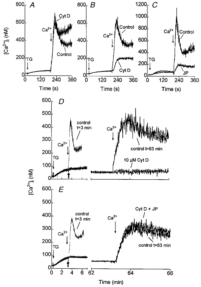
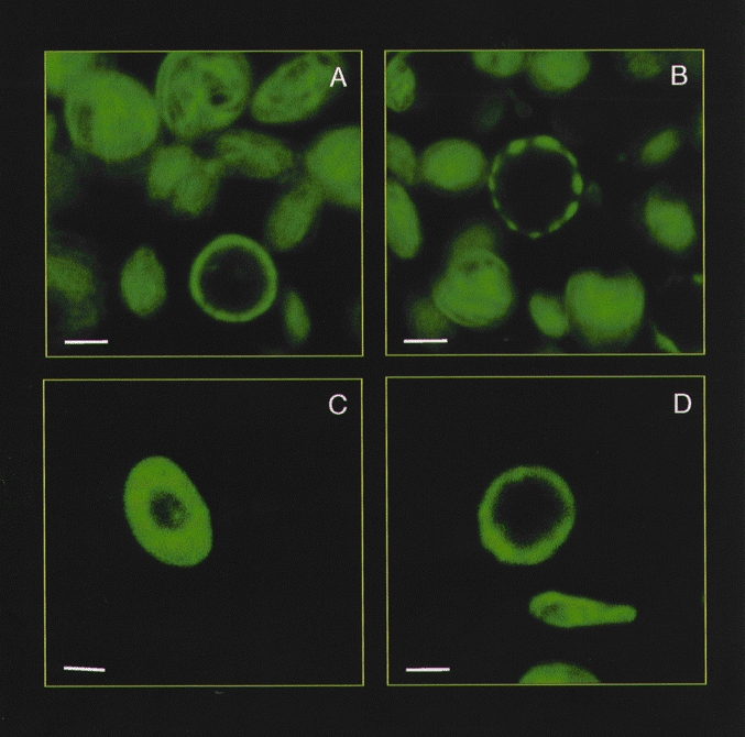
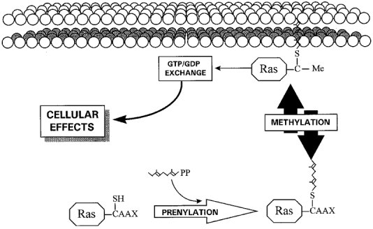
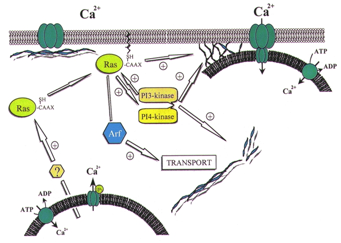
Store-mediated Ca2+ entry is the main pathway for Ca2+ influx in platelets and many other cells. Several hypotheses have considered both direct and indirect coupling mechanisms between the endoplasmic reticulum and the plasma membrane. Here we pay particular attention to new insights into the regulation of store-mediated Ca2+ entry: the role of the cytoskeleton in a secretion-like coupling model. In this model, Ca2+ entry may be mediated by a reversible trafficking and coupling of the endoplasmic reticulum with the plasma membrane, that shows close parallels to the events mediating secretion. As with secretion, the actin cytoskeleton plays an inhibitory role in the activation of Ca2+ entry by preventing the approach and coupling of the endoplasmic reticulum with the plasma membrane, making cytoskeletal remodelling a key event in the activation of Ca2+ entry. We also review recent advances investigating the regulation of store-mediated Ca2+ entry by small GTPases and phosphoinositides, which might be involved in the store-mediated Ca2+ entry pathway through roles in the remodelling of the cytoskeleton.


Figures

Figure 1
Figure 1. Effects of cytoskeletal modifiers on SMCE in human platelets
Fura-2-loaded human platelets were incubated at 37 °C either in the presence of 10 μm cytochalasin D (Cyt D) for 1 min (A) or 40 min (B), or in the presence of 10 μm jasplakinolide (JP) for 30 min (C) or the vehicles as controls. At the time of experiment 100 μm EGTA was added. Cells were then stimulated with 200 nM thapsigargin (TG) and 3 min later CaCl2 (final concentration 300 μm) was added to the medium to initiate Ca2+ entry. D and E, human platelets suspended in a Ca2+-free medium were stimulated with TG (200 nM) and 3 min later 10 μm Cyt D (D), 10 μm Cyt D plus 10 μm JP (E), or the vehicle (control t= 63 min) were added as indicated by the thick arrows. CaCl2 (300 μm) was added to the medium at the same time, as a control (control t= 3 min) or 1 h after Cyt D, Cyt D plus JP or the vehicle to initiate Ca2+ entry. Adapted from Rosado et al. (2000).
Figure 2
Figure 2. Effect of cytoskeletal modifiers on the reorganisation of the actin cytoskeleton in human platelets
Confocal microscopy of human platelets stained with FITC-labelled phalloidin, an actin-binding fungal toxin. A, control conditions. B, platelets treated with 10 μm cytochalasin D for 40 min. C, platelets treated with 10 μm jasplakinolide for 30 min. D, platelets treated with 100 μm calyculin A for 2 min. The bars represent 1 μm.
Figure 3
Figure 3. Overview of prenylation and methylation of Ras proteins
Modification of the C-terminus cysteine residue (designated ‘C’) by attachment of a farnesyl or geranylgeranyl group, followed by the removal of the three C-terminal amino acids and reversible methylation. ‘C-Me’ indicates that the carboxyl group has been methylated.
Figure 4
Figure 4. Proposed secretion-like coupling model for SMCE in human platelets
Depletion of the intracellular Ca2+ stores induces translocation and association of the small GTP-binding proteins of the Ras superfamily with the plasma membrane. Association of Ras proteins with the plasma membrane facilitates their interaction with GDP/GTP exchangers and, therefore, their activation. Ras proteins promote actin polymerisation leading the trafficking of the endoplasmic reticulum towards the plasma membrane. In addition, Arf, a member of the Ras small GTP-binding proteins, is involved in intracellular transport. Ras proteins induce reorganisation of the membrane actin filaments to support the coupling between elements in the endoplasmic reticulum (possibly the InsP3 receptors: ‘IP3’ in figure) and the Ca2+ release-activated channel (CRAC) in the plasma membrane through a protein-protein interaction. The effect of Ras proteins might be mediated to some extent by the activation of phosphoinositide 3′ and 4′ kinases, which in turn promote actin cytoskeleton rearrangement and activation of different Ras proteins.

References

    1. Álvarez J, Montero M, García-Sancho J. Cytochrome P450 may regulate plasma membrane Ca2+ permeability according to the filling state of the intracellular Ca2+ stores. FASEB Journal. 1992;6:786–792. - PubMed
    1. Ando S, Kaibuchi K, Sasaki T, Hiraoka K, Nishiyama T, Mizuno T, Asada M, Nunoi H, Matsuda I, Matsuura Y, Polakis P, McCormick F, Takai Y. Post-translational processing of rac p21s is important both for their interaction with the GDP/GTP exchange proteins and for their activation of NADPH oxidase. Journal of Biological Chemistry. 1992;267:25709–25713. - PubMed
    1. Berridge MJ. Capacitative calcium entry. Biochemical Journal. 1995;312:1–11. - PMC - PubMed
    1. Bird GStJ, Putney JW., Jr Inhibition of thapsigargin-induced calcium entry by microinjected guanine nucleotide analogues. Evidence for the involvement of a small G-protein in capacitative calcium entry. Journal of Biological Chemistry. 1993;268:21486–21488. - PubMed
    1. Brown BL, Walker SW, Tomlinson S. Calcium calmodulin and hormone secretion. Clinical Endocrinology. 1985;23:201–218. - PubMed

Publication types

LinkOut - more resources