. 2000 Jul 17;19(14):3556-64.
doi: 10.1093/emboj/19.14.3556.

Vacuolar uptake of host components, and a role for cholesterol and sphingomyelin in malarial infection

Affiliations

Vacuolar uptake of host components, and a role for cholesterol and sphingomyelin in malarial infection

S Lauer et al. EMBO J. .

Abstract

Erythrocytes, which are incapable of endocytosis or phagocytosis, can be infected by the malaria parasite Plasmodium falciparum. We find that a transmembrane protein (Duffy), glycosylphosphatidylinositol (GPI)-anchored and cytoplasmic proteins, associated with detergent-resistant membranes (DRMs) that are characteristic of microdomains in host cell membranes, are internalized by vacuolar parasites, while the major integral membrane and cytoskeletal proteins are not. The internalized host proteins and a plasmodial transmembrane resident parasitophorous vacuolar membrane (PVM) protein are detected in DRMs associated with vacuolar parasites. This is the first report of a host transmembrane protein being recruited into an apicomplexan vacuole and of the presence of vacuolar DRMs; it establishes that integral association does not preclude protein internalization into the P.FALCIPARUM: vacuole. Rather, as shown for Duffy, intracellular accumulation occurs at the same rate as that seen for a DRM-associated GPI-anchored protein. Furthermore, novel mechanisms regulated by the DRM lipids, sphingomyelin and cholesterol, mediate (i) the uptake of host DRM proteins and (ii) maintenance of the intracellular vacuole in the non-endocytic red cell, which may have implications for intracellular parasitism and pathogenesis.


Figures
None
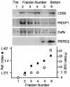
Fig. 1. Density gradient detection of DRMs from uninfected erythrocytes. Cold Triton X-100 extracts were prepared, subjected to sucrose density gradients, and the resulting fractions were analyzed by western blotting and densitometric scanning (using Molecular Dynamics ImageQuant 5.0 software) for the distribution of indicated markers (see Materials and methods). Data from one representative experiment are shown. Floating fractions 2–4 indicate DRMs.
None
Fig. 2. Internalization of host DRM proteins in infected erythrocytes. Infected red cells were probed with primary and relevant secondary antibodies in i–v and vii–xii to detect the indicated markers. (i) Non-immune; (ii–v) CD59, red; (iv) PfEXP1, green; (v) BODIPY-ceramide, green; (vi) FITC–anti-CD59, green; (vii) CD55; (viii) band 3; (ix) glycophorin A; (x and xi) Duffy, green; (xii) Gαs, green. No internalization of CD59 was seen in uninfected red cells and western blots confirmed that the antibody recognizes a single ∼19 kDa protein present at equal levels in both infected and uninfected cells (data not shown). No internalization of Duffy or Gαs proteins was seen in uninfected cells and antibodies to these proteins recognized only their respective host polypeptides in both infected and uninfected cells (data not shown). In (vi), infected red cells were labeled with anti-CD59 antibody that was directly conjugated to FITC. In all images, the nucleus (blue) is stained with Hoechst, v indicates the periphery of the red cell, arrows indicate vacuolar parasite, asterisks indicate TVM and the scale bar is 5 µm.
None
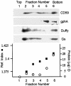
Fig. 3. Density gradient detection of DRMs from isolated vacuolar parasites. Vacuolar parasites were isolated and the level of red cell contamination judged by band 3 and gph A staining was <5% (see Materials and methods). Fractions were obtained (as described in Materials and methods and Figure 1) and analyzed for the distribution of indicated markers. Data from one experiment are shown. Floating fractions 2–4 indicate DRMs.
None
Fig. 4. Internalization of host DRM lipids in infected red cells. (A) Cholesterol in a (i) ring and (ii) trophozoite as detected by filipin staining (Haldar et al., 1991). (B) GL4 in (i) rings and (ii) schizont as detected in an indirect immunofluorescence assay using anti-GL4 and FITC-conjugated secondary antibody.
None
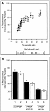
Fig. 5. Quantitative uptake of CD59 and Duffy and their inhibition by PPMP in infected red cells. (A) Amount of cell-associated CD59 and Duffy detected in association with the vacuolar parasite as a function of size and corresponding hours of intracellular parasite development. CD59 (filled squares) and Duffy (open circles) fluorescence associated with the parasite and red cell were quantitated as described in Materials and methods. Each point represents an average of 10 P.falciparum-infected red cells. (B) Vacuolar accumulation of CD59 (black bars) and Duffy (open bars) in: (lanes 1 and 2) mock-treated cells at 36 h of development; (lanes 3 and 4) 24 h trophozoites incubated with 5 µM PPMP for a subsequent 12 h in culture; (lanes 5 and 6) 0–6 h rings exposed to 5 µM PPMP for the next 30 h in culture.
None
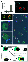
Fig. 6. Effects of cholesterol depletion on intracellular trophozoite release. (A) Fraction of parasites released when cells were treated with (1) RPMI, (2) RPMI + cyclodextrin–cholesterol complex and (3) RPMI + cyclodextrin. (B) Cyclodextrin-treated cells were fixed in 1% glutaraldehyde and stained with filipin (blue) and ethidium bromide (red) and viewed by fluorescence microscopy. v indicates an uninfected cell; arrows indicate parasites. Scale bar is 5 µm. (C) Indirect immunofluorescence assay of (i) mock- and (ii–iv) cyclodextrin-treated cells. In (i) and (ii), cells were stained for the PPM marker PfMSP1 (green) and the PVM marker PfEXP1 (red). Arrows indicate ‘spots’ of PfEXP1 detected at one end of the parasite or in association with red cell ghosts (negative contrast not shown). In (iii) and (iv), cells were labeled with a red cell marker band 3 (green) and the PVM marker PfEXP1 (red). Blue indicates Hoechst-stained nuclei. Scale bar indicates 5 µm. (D) Summary of the association of the PVM and the PPM under different conditions of parasite release from red cells. Depletion of erythrocyte surface cholesterol releases parasites freed of their surrounding vacuolar membrane. Other known methods of parasite release, such as permeabilization with digitonin/saponin or mechanical homogenization, fail to separate vacuole from the parasite.

References

    1. Anderson R. (1998) The caveolae membrane system. Annu. Rev. Biochem., 67, 199–225. - PubMed
    1. Barnwell J.W. (1990) Vesicle-mediated transport of membrane and proteins in malaria-infected erythrocytes. Blood Cells, 16, 379–395. - PubMed
    1. Brown D.A. and London,E. (1998a) Functions of lipid rafts in biological membranes. Annu. Rev. Cell Dev. Biol., 14, 111–136. - PubMed
    1. Brown D.A. and London,E. (1998b) Structure and origin of ordered lipid domains in biological membranes. J. Membr. Biol., 164, 103–114. - PubMed
    1. Brown D.A. and Rose,J.,K. (1992) Sorting of GPI-anchored proteins to glycolipid-enriched membrane subdomains during transport to the apical cell surface. Cell, 68, 533–544. - PubMed

Publication types

MeSH terms