. 2000 Jul 17;19(14):3704-13.
doi: 10.1093/emboj/19.14.3704.

A dual role for zinc fingers in both DNA binding and zinc sensing by the Zap1 transcriptional activator

Affiliations

A dual role for zinc fingers in both DNA binding and zinc sensing by the Zap1 transcriptional activator

A J Bird et al. EMBO J. .

Abstract

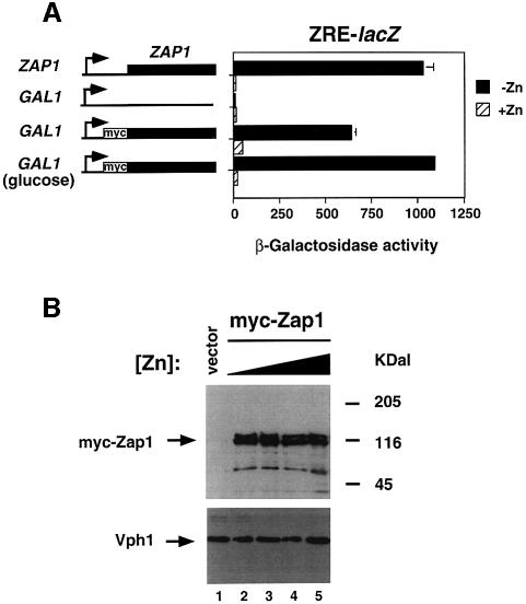
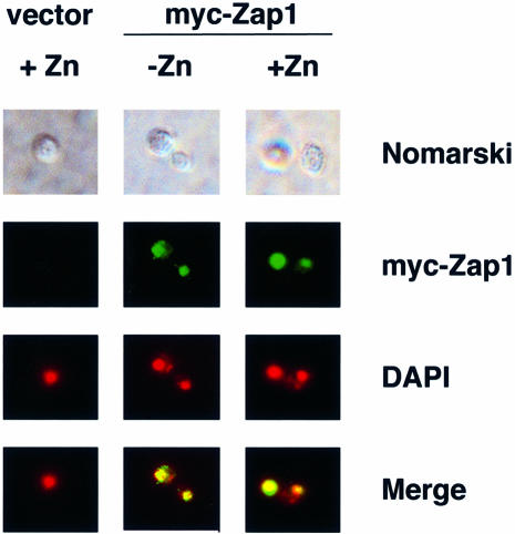
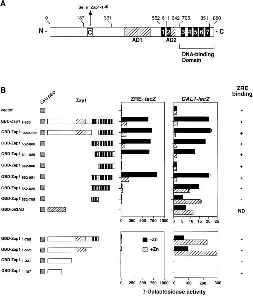
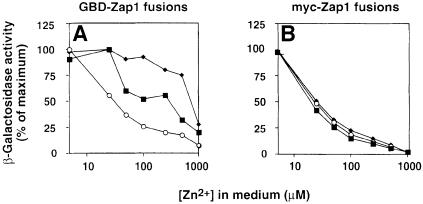
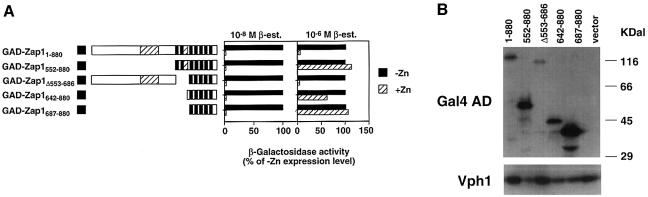
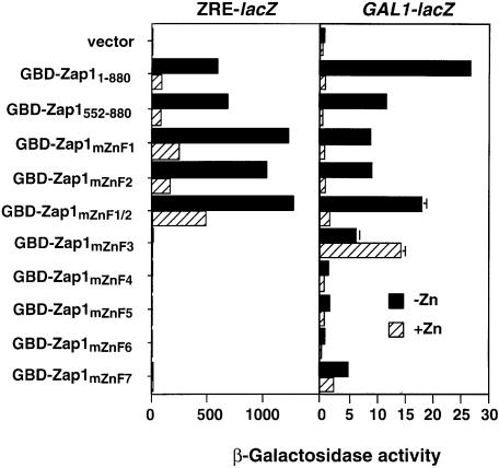
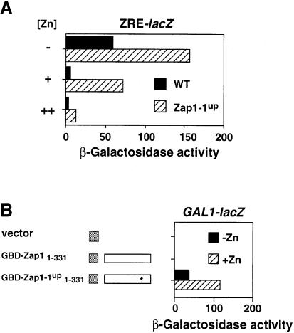
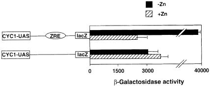
The Zap1 transcriptional activator of Saccharomyces cerevisiae controls zinc homeostasis. Zap1 induces target gene expression in zinc-limited cells and is repressed by high zinc. One such target gene is ZAP1 itself. In this report, we examine how zinc regulates Zap1 function. First, we show that transcriptional autoregulation of Zap1 is a minor component of zinc responsiveness; most regulation of Zap1 activity occurs post-translationally. Secondly, nuclear localization of Zap1 does not change in response to zinc, suggesting that zinc regulates DNA binding and/or activation domain function. To understand how Zap1 responds to zinc, we performed a functional dissection of the protein. Zap1 contains two activation domains. DNA-binding activity is conferred by five C-terminal C(2)H(2) zinc fingers and each finger is required for high-affinity DNA binding. The zinc-responsive domain of Zap1 also maps to the C-terminal zinc fingers. Furthermore, mutations that disrupt some of these fingers cause constitutive activity of a bifunctional Gal4 DNA-binding domain-Zap1 fusion protein. These results demonstrate a novel function of Zap1 zinc fingers in zinc sensing as well as DNA binding.


Figures

None
Fig. 1. Regulation of Zap1 activity by zinc occurs at a post-translational level. (A) Wild-type (DY1457) and zap1 mutant (ZHY6) cells containing either the pYef2 vector or pMyc-Zap11–880 were grown to exponential phase in LZM-galactose supplemented with either 5 µM (–Zn) or 1000 µM (+Zn) ZnCl2. Zap1 activity in each strain was assessed using the pDg2 ZRE-lacZ reporter. ZHY6 pMyc-Zap11–880 transformants were also assayed after growth in glucose, a carbon source that represses most but not all expression from the GAL1 promoter. A representative experiment is shown and the error bars indicate 1 SD. (B) The stability of Zap1 is not affected by zinc status. Wild-type (DY1457) cells transformed with the pYef2 vector and zap1 mutant (ZHY6) cells bearing pMyc-Zap11–880 were grown in LZM-galactose to exponential phase. The concentrations of ZnCl2 added to the medium were 5 (lane 2), 250 (lane 3), 500 (lane 4) and 1000 µM (lanes 1 and 5). Crude protein extracts were prepared, fractionated by SDS–PAGE analysis, and assayed for Zap1 and Vph1 protein levels by immunoblotting.
None
Fig. 2. The subcellular localization of Zap1 is not affected by zinc status. Wild-type (DY1457) cells bearing the vector pYef2 and zap1 mutant (ZHY6) cells bearing pMyc-Zap11–880 were grown to exponential phase in LZM-galactose supplemented with either 5 µM (–Zn) or 1000 µM (+Zn) ZnCl2. Cells were viewed by Nomarski optics or epifluorescence. DAPI was used to stain the nucleus and the myc-Zap1 protein was detected by indirect immunofluorescence. The blue fluorescence of DAPI staining was converted to red and the DAPI and myc-Zap1 images were overlaid using Adobe Photoshop (merge). Yellow color in the merged images indicates colocalization of the markers.
None
Fig. 3. The functional domains of Zap1. (A) Schematic representation of Zap1. Shown are the functional activation domains (AD1 and AD2, hatched boxes) and the DNA-binding domain. The seven putative zinc finger domains are represented by black boxes and are numbered 1–7. Amino acid positions relevant to the constructed plasmid fusions are also numbered. The position of the Cys→Ser substitution in the Zap1-1up protein is shown. (B) Mapping the ZRD of Zap1. The activity of GBD–Zap1 fusion proteins expressed from the ADH1 promoter in the strain DEY1538 (zap1 gal4 gal80) was measured using either a ZRE-lacZ (pDg2) or a GAL1-lacZ (pRY171) reporter. Cells were grown in LZM supplemented with either 5 µM (–Zn) or 1000 µM (+Zn) ZnCl2 prior to the β-galactosidase activity assays. A representative experiment is shown and the error bars indicate 1 SD. Accumulation of all fusion proteins was confirmed by immunoblot analysis using an anti-GBD antibody. EMSA with a ZRE oligonucleotide probe (Zhao et al., 1998) was used to assess DNA-binding activity. ND, not determined.
None
Fig. 4. Comparison of GBD–Zap1 and myc-Zap1 activities in response to a range of zinc concentrations. (A) A zap1 mutant strain (ZHY6) bearing the pDg2 ZRE-lacZ reporter and pGBD–Zap11–880 (filled squares), pGBD–Zap1Δ553–686 (filled diamonds) or pGBD–Zap1552–880 (open circles) was grown to exponential phase in LZM supplemented with the indicated concentrations of ZnCl2 prior to assay for β-galactosidase activity. (B) The same zap1 mutant strain bearing the pDg2 ZRE-lacZ reporter and pMyc-Zap11–880 (filled squares), pMyc-Zap1Δ553–686 (filled diamonds) or pMyc-Zap1552–880 (open circles) was grown to exponential phase in LZM supplemented with the indicated concentrations of ZnCl2 prior to assay for β-galactosidase activity. Shown are representative experiments in which the standard deviations were <10% of the corresponding mean.
None
Fig. 5. Mapping the ZRD with GAD fusions. The indicated fusions were expressed in a zap1 gal4 gal80 mutant strain (DEY1538) bearing the pDg2 ZRE-lacZ reporter and pGEV-HIS3. pGEV-HIS3 encodes a hybrid transcriptional activator that is induced by β-estradiol. (A) Cells were grown to exponential phase in LZM supplemented with either 5 µM (–Zn) or 1000 µM (+Zn) ZnCl2 and either 10–8 or 10–6 M β-estradiol. β-galactosidase activities are represented as a percentage of the corresponding –Zn expression level and the standard deviations were <10% of the corresponding mean. (B) The same transformants as in (A) were grown to exponential phase in LZM supplemented with 1000 µM ZnCl2; protein extracts were prepared and analyzed by immunoblotting using anti-GAD or anti-Vph1 antibodies. Apparent proteolytic products were observed in some samples; i.e. the largest band in each lane corresponds to the expected molecular mass of each fusion.
None
Fig. 6. Effects of zinc finger mutations on zinc responsiveness. A zap1 gal4 gal80 mutant strain (DEY1538) containing GBD–Zap11–880, GBD–Zap1552–880 or GBD–Zap1mZnFX (where X indicates the mutated zinc finger domain) was grown in LZM supplemented with either 5 µM (–Zn) or 1000 µM (+Zn) ZnCl2. β-galactosidase activity was measured in each strain using either a ZRE-lacZ (pDg2) or a GAL1-lacZ (pRY171) reporter. A representative experiment is shown and the error bars indicate 1 SD.
None
Fig. 7. The ZAP1-1up mutation turns on a quiescent activation domain. (AZAP1-1up retains zinc-responsive regulation. Wild-type (DY1457) and ZAP1-1up (ZHY7) strains bearing pDg2 were grown to exponential phase in SD media (+) or in SD media that had been supplemented with 1 mM EDTA, 100 µM ZnCl2 (–) or 100 µM ZnCl2 alone (++) prior to β-galactosidase activity assays. (B) The ZAP1-1up allele contains an activation domain not present in the wild-type protein. Strain DEY1538 (zap1 gal4 gal80) containing the GAL1-lacZ reporter (pRY171) and either the vector (pMA424), pGBD–Zap11–331 or pGBD Zap1-1up1–331 was grown to exponential phase in LZM supplemented with either 5 µM (–Zn) or 1000 µM (+Zn) ZnCl2 prior to β-galactosidase assay. The asterisk denotes the mutation in the Zap1-1up allele. Representative experiments are shown.
None
Fig. 8. Zap1 activity in zinc-replete cells is not repressed by a repressor capable of acting on nearby UASs. Wild-type (DY1457) cells transformed with pLGΔ312 or pZRE-LGΔ312 were grown to exponential phase in LZM supplemented with either 5 µM (–Zn) or 1000 µM (+Zn) ZnCl2 prior to β-galactosidase assay. A representative experiment is shown and the error bars indicate 1 SD.

References

    1. Baeuerle P.A. and Baltimore,D. (1988) IκB: a specific inhibitor of the NF-κB transcription factor. Science, 242, 540–545. - PubMed
    1. Bird A.J., Evans-Galea,M., Blankman,E., Zhao,H., Luo,H., Winge,D.R. and Eide,D.J. (2000) Mapping the DNA binding domain of the Zap1 zinc-responsive transcriptional activator. J. Biol. Chem., 275, 16160–16166. - PubMed
    1. Bradford M.M. (1976) A rapid and sensitive method for the quantitation of microgram quantities of protein utilizing the principle of protein-dye binding. Anal. Biochem., 72, 248–254. - PubMed
    1. Cross F.R. (1997) ‘Marker swap’ plasmids: convenient tools for budding yeast molecular genetics. Yeast, 13, 647–663. - PubMed
    1. Cullin C. and Minvielle-Sebastia,L. (1994) Multipurpose vectors designed for the fast generation of N- or C-terminal epitope-tagged proteins. Yeast, 10, 105–112. - PubMed

Publication types

MeSH terms