. 2000 Jul 17;19(14):3831-40.
doi: 10.1093/emboj/19.14.3831.

Telomere-led bouquet formation facilitates homologous chromosome pairing and restricts ectopic interaction in fission yeast meiosis

Affiliations

Telomere-led bouquet formation facilitates homologous chromosome pairing and restricts ectopic interaction in fission yeast meiosis

O Niwa et al. EMBO J. .

Abstract

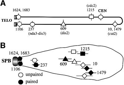
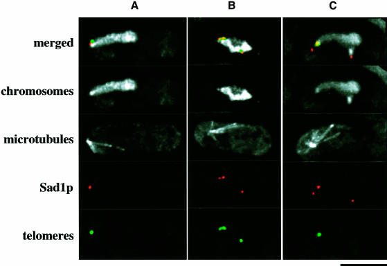
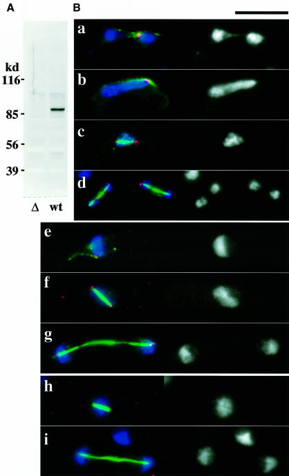
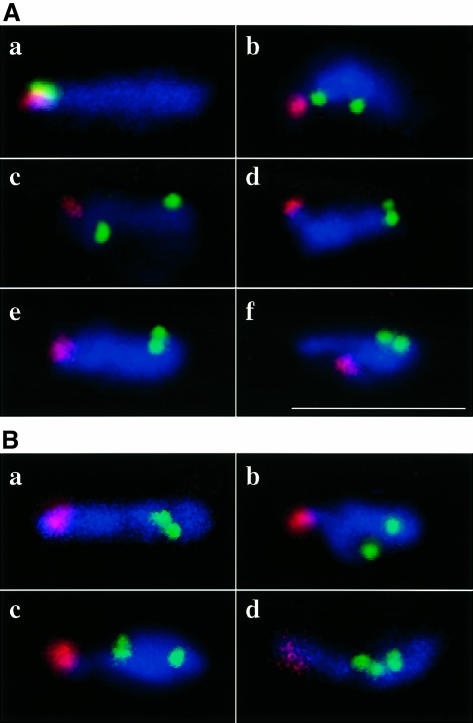
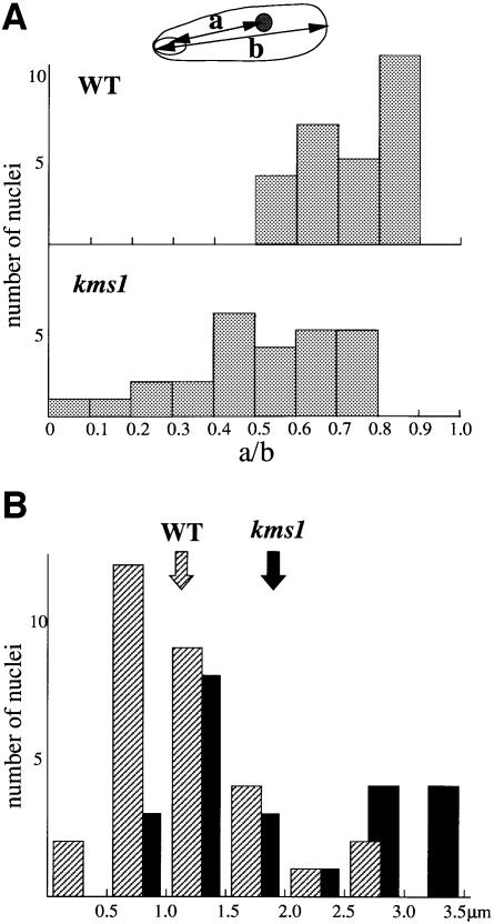
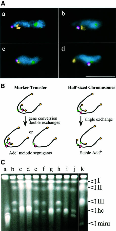
A polarized chromosomal arrangement with clustered telomeres in a meiotic prophase nucleus is often called bouquet and is thought to be important for the pairing of homologous chromosomes. Fluorescence in situ hybridization in fission yeast indicated that chromosomal loci are positioned in an ordered manner as anticipated from the bouquet arrangement. Blocking the formation of the telomere cluster with the kms1 mutation created a disorganized chromosomal arrangement, not only for the regions proximal to the telomere but also for interstitial regions. The kms1 mutation also affected the positioning of a linear minichromosome. Consistent with this cytological observation, the frequency of ectopic homologous recombination between a linear minichromosome and a normal chromosome increased in the kms1 background. Intragenic recombination between allelic loci is reduced in the kms1 mutant, but those between non-allelic loci are unaffected or slightly increased. Thus, telomere-led chromosome organization facilitates homologous pairing and also restricts irregular chromosome pairing during meiosis.


Figures

None
Fig. 1. (A) A physical map of cosmids used in this study. The cosmid number is as described in Mizukami et al. (1993). Marker genes carried by a respective cosmid are shown in parentheses. (B) Positioning of each cosmid sequence in the meiotic prophase nucleus as revealed using FISH. Horizontal bars indicate the standard deviation.
None
Fig. 2. Multiple Sad1 stains observed in meiotic prophase nuclei in the kms1 mutant. Chromosomes, microtubules, Sad1p and telomeres were stained with DAPI, TAT1, anti-Sad1 and cos212 DNA probe, respectively. Merged images, chromosomes and mictrotubules: white; Sad1p: red; telomeres: green. (A) Wild type, (B and C) kms1-1 mutant. Scale bar, 5 µm.
None
Fig. 3. (A) Specificity of the anti-Kms1 antibody. Immunoblot analysis of a kms1-disrupted (left lane) and a wild-type (right) cell extract with the antibody. (B) Localization of the kms1+ product. (a–g) Wild type; (h and i) cells with the disrupted kms1. See the text for details. In each panel, the left side is a merged image of chromosomes (blue), microtubules (green) and Kms1p (red). The right hand side shows chromosomes stained with DAPI. Scale bar, 10 µm.
None
Fig. 4. (A) FISH analysis of wild type (a) and kms1 mutant (b–f) meiotic prophase nuclei. Red, rDNA; green, cos1683. Nuclei are stained with DAPI (blue). (B) Wild type (a) and kms1 mutant (b–d). Red, rDNA; green, cos1479; blue, DAPI. In (c and d), prematurely separated sister chromatids are shown. Scale bar, 10 µm.
None
Fig. 5. (A) Positioning of the centromere-proximal sequence in the meiotic nuclei. (a) The distance from the center of the rDNA signal to the center of the cos1479 signal; (b) the length of the respective nucleus. (B) Scattering of centromeres in the meiotic nuclei. Distribution of the longest distance between pairs of centromeres in individual zygote. Shaded bars, wild type; solid bars, kms1. The arrows indicate average distances.
None
Fig. 6. (A) FISH analysis of wild-type (a) and kms1 mutant (b–d) meiotic prophase nuclei containing the minichromosome ChR33-Tr29. Yellow, rDNA (a, b and d) or telomeres (cos212 in c); green, centromeres (pRS140); red, minichromosome (YIp32). Note that the minichromosome also contains the pRS140 sequence. Nuclei are stained with DAPI (cyan). Scale bar, 10 µm. (B) Experimental designs for the detection of meiotic recombination between a minichromosome and chromosome III (see the text). The rectangles represent the Ade marker. (C) Pulsed-field gel electrophoresis of stable Ade+ segregants (see the text for details). (Lane a) Chromosomes of S.cerevisiae; (lanes b–j) Ade+ segregants from the mutant cross; (lane k) S.pombe strain containing Ch16 minichromosome. The positions of chromosomes I, II and III, half-sized chromosomes (HC), and minichromosome Ch16 (mini) are indicated on the right hand side.

References

    1. Alfa C., Fantes,P., Hyams,J., McLeod,M. and Warbrick,E. (1993) Experiments With Fission Yeast: A Laboratory Course Manual. Cold Spring Harbor Laboratory Press, Cold Spring Harbor, NY.
    1. Allshire R.C. (1992) Manipulation of large minichromosomes in Schizosaccharomyces pombe with liposome-enhanced transform ation. Methods Enzymol., 216, 614–631. - PubMed
    1. Bahler J., Wyler,T., Loidl J. and Kohli,J. (1993) Unusual nuclear structures in meiotic prophase of fission yeast: a cytological analysis. J. Cell Biol., 121, 241–256. - PMC - PubMed
    1. Bass H.W., Marshall,W.F., Sedat,J.W., Agard,D.A. and Cande,W.Z. (1997) Telomere cluster de novo before the initiation of synapsis: a three-dimensional spatial analysis of telomere positions before and during meiotic prophase. J. Cell Biol., 137, 5–18. - PMC - PubMed
    1. Chikashige Y., Ding,D.-Q., Funabiki,H., Haraguchi,T., Mashiko,S., Yanagida,M. and Hiraoka,Y. (1994) Telomere-led premeiotic chromosome movement in fission yeast. Science, 264, 270–273. - PubMed

Publication types

MeSH terms

Substances