. 2000 Aug;68(8):4391-8.
doi: 10.1128/IAI.68.8.4391-4398.2000.

beta-1,2-linked oligomannosides from Candida albicans bind to a 32-kilodalton macrophage membrane protein homologous to the mammalian lectin galectin-3

Affiliations

beta-1,2-linked oligomannosides from Candida albicans bind to a 32-kilodalton macrophage membrane protein homologous to the mammalian lectin galectin-3

C Fradin et al. Infect Immun. 2000 Aug.

Abstract

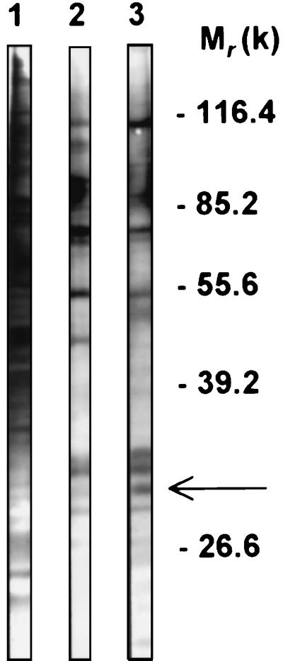
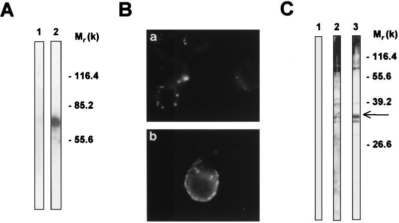
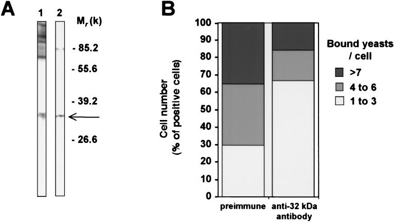
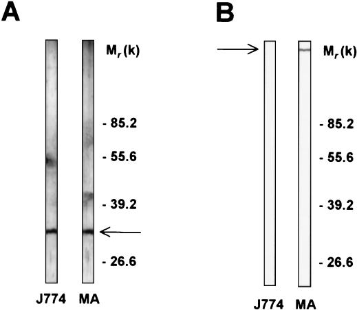
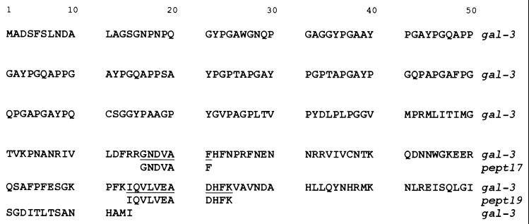
beta-1,2-linked oligomannoside residues are present, associated with mannan and a glycolipid, the phospholipomannan, at the Candida albicans cell wall surface. beta-1,2-linked oligomannoside residues act as adhesins for macrophages and stimulate these cells to undergo cytokine production. To characterize the macrophage receptor involved in the recognition of C. albicans beta-1,2-oligomannoside we used the J774 mouse cell line, which is devoid of the receptor specific for alpha-linked mannose residues. A series of experiments based on affinity binding on either C. albicans yeast cells or beta-1,2-oligomannoside-conjugated bovine serum albumin (BSA) and subsequent disclosure with biotinylated conjugated BSA repeatedly led to the detection of a 32-kDa macrophage protein. An antiserum specific for this 32-kDa protein inhibited C. albicans binding to macrophages and was used to immunoprecipitate the molecule. Two high-pressure liquid chromatography-purified peptides from the 32-kDa tryptic digest showed complete homology to galectin-3 (previously designated Mac-2 antigen), an endogenous lectin with pleiotropic functions which is expressed in a wide variety of cell types with which C. albicans interacts as a saprophyte or a parasite.


Figures

FIG. 1
FIG. 1
Binding analysis of J774 surface membrane proteins to yeast cells. Plasma membrane proteins from J774 cells were biotinylated and extracted using Triton X-100. Soluble extracts were incubated with either S. cerevisiae (lane 2) or C. albicans (lane 3) blastoconidia. After the samples were washed, the bound proteins were eluted, resolved by SDS-PAGE, and subjected to blotting with alkaline phosphatase-conjugated streptavidin. Lane 1 contains total extract from biotinylated cells. The arrow indicates the major protein differing between the two yeast-associated proteins. Molecular weights (in thousands) are shown on the right.
FIG. 2
FIG. 2
Identification and localization of the 32-kDa macrophage protein recognized by β-1,2-Man–BSA. (A) After conjugation, the presence of β-1,2-oligomannosidic epitopes on biot-β-1,2-Man–BSA was examined using Western blotting with anti-β-1,2-oligomannoside MAb AF1 (lane 2). Lane 1 shows the reactivity of MAb AF1 with uncoupled BSA. (B) J774 cells were incubated with biot-BSA (a) or biot-β-1,2-Man–BSA (b). Bound protein was revealed with fluorescein-conjugated streptavidin. (C) Extracts from J774 cells were subjected to blotting with biot-BSA (lane 1), biot-α-Man–BSA (lane 2), or biot-β-1,2-Man–BSA (lane 3) and revealed with peroxidase-conjugated streptavidin. The arrow indicates the position of the 32-kDa protein. These experiments were performed three to five times with identical results. Molecular weights (in thousands) are given on the right of panels A and C.
FIG. 3
FIG. 3
Comparison of macrophage proteins recognized by β-1,2-Man–BSA and C. albicans blastoconidia. J774 extracts were incubated with C. albicans yeast cells (A) or with Sepharose beads coupled with β-1,2-Man–BSA (B). Bound material was eluted, electrophoresed, and blotted with biot-BSA (lanes 1) or biot-β-1,2-Man–BSA (lane 2). The arrows indicate the position of the 32-kDa protein. Experiments were repeated at least five times with identical results. Molecular weights (in thousands) are given on the right of the panels.
FIG. 4
FIG. 4
Specificity of the anti-32-kDa polyclonal antibody that inhibits C. albicans binding to J774 cells. (A) J774 proteins eluted from C. albicans blastoconidia were revealed either with biot-β-1,2-Man–BSA (lane 1) or with the rat polyclonal antibody raised against the 32-kDa protein (lane 2). (B) J774 cells (105 cells per well) were cultured at 37°C in eight-well Lab-Tek tissue culture chambers in the presence of a 1:100 dilution of either preimmune or immune anti-32 kDa polyclonal antibody. C. albicans yeast cells were then added at a yeast-to-cell ratio of 50:1. The slides were washed, fixed, and mounted for microscopy examination. Data are expressed as the number of J774 cells in relation to the number of yeasts ingested by cell. Experiments were repeated at least five times with similar results. Molecular weights (in thousands) are given on the right of panel A.
FIG. 5
FIG. 5
Comparison of antigenicity of the 32-kDa protein and the MMR. Extracts of mouse peritoneal macrophages (MA) or from J774 cells (J774) were blotted with either anti-32-kDa (A) or anti-MMR (B) polyclonal antibodies and revealed with specific antiserum conjugated to peroxidase. Experiments were repeated at least three times with identical results.
FIG. 6
FIG. 6
Sequence homologies between two peptides of the 32-kDa protein and galectin-3. The J774 cell 32-kDa protein was immunoprecipitated with the anti-32-kDa antiserum, resolved by SDS-PAGE, and subjected to trypsin digestion as described in Materials and Methods. Peptides were separated by HPLC. Homologies of two peptides (pept17 and pept19) to known proteins were examined with the Swiss-Prot database.

References

    1. Barondes S H, Cooper D N, Gitt M A, Leffler H. Galectins. Structure and function of a large family of animal lectins. J Biol Chem. 1994;269:20807–20810. - PubMed
    1. Cassone A, Torosantucci A, Boccanera M, Pelligrini G, Palma C, Malavasi F. Production and characterization of a monoclonal antibody to a cell-surface, glucomannoprotein constituent of Candida albicans and other pathogenic Candida species. J Med Microbiol. 1988;27:233–238. - PubMed
    1. Castro M, Ralston N V, Morgenthaler T I, Rohrbach M S, Limper A H. Candida albicans stimulates arachidonic acid liberation from alveolar macrophages through alpha-mannan and beta-glucan cell wall components. Infect Immun. 1994;62:3138–3145. - PMC - PubMed
    1. Csank C, Schroppel K, Leberer E, Harcus D, Mohamed O, Meloche S, Thomas D Y, Whiteway M. Roles of the Candida albicans mitogen-activated protein kinase homolog, Cek1p, in hyphal development and systemic candidiasis. Infect Immun. 1998;66:2713–2721. - PMC - PubMed
    1. Czop J K, Austen K F. A beta-glucan inhibitable receptor on human monocytes: its identity with the phagocytic receptor for particulate activators of the alternative complement pathway. J Immunol. 1985;134:2588–2593. - PubMed

Publication types

LinkOut - more resources