

Invasion of epithelial cells by Yersinia pestis: evidence for a Y. pestis-specific invasin
- PMID: 10899851
- PMCID: PMC98364
- DOI: 10.1128/IAI.68.8.4523-4530.2000
Invasion of epithelial cells by Yersinia pestis: evidence for a Y. pestis-specific invasin
Abstract
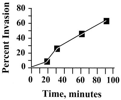
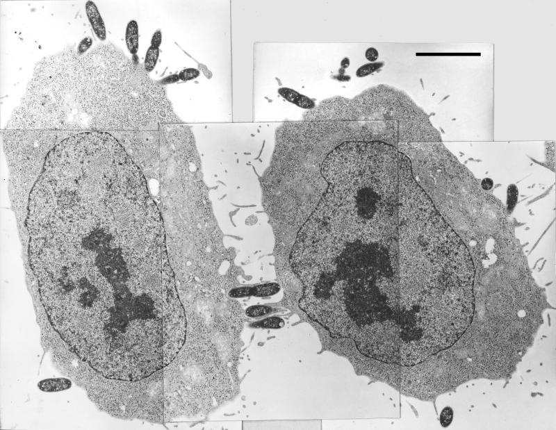
The causative agent of plague, Yersinia pestis, is regarded as being noninvasive for epithelial cells and lacks the major adhesins and invasins of its enteropathogenic relatives Yersinia enterocolitica and Yersinia pseudotuberculosis. However, there are studies indicating that Y. pestis invades and causes systemic infection from ingestive and aerogenic routes of infection. Accordingly, we developed a gentamicin protection assay and reexamined invasiveness of Y. pestis for HeLa cells. By optimizing this assay, we discovered that Y. pestis is highly invasive. Several factors, including the presence of fetal bovine serum, the configuration of the tissue culture plate, the temperature at which the bacteria are grown, and the presence of the plasminogen activator protease Pla-encoding plasmid pPCP1, were found to influence invasiveness strongly. Suboptimal combinations of these factors may have contributed to negative findings by previous studies attempting to demonstrate invasion by Y. pestis. Invasion of HeLa cells was strongly inhibited by cytochalasin D and modestly inhibited by colchicine, indicating strong and modest respective requirements for microfilaments and microtubules. We found no significant effect of the iron status of yersiniae or of the pigmentation locus on invasion and likewise no significant effect of the Yops regulon. However, an unidentified thermally induced property (possibly the Y. pestis-specific capsular protein Caf1) did inhibit invasiveness significantly, and the plasmid pPCP1, unique to Y. pestis, was essential for highly efficient invasion. pPCP1 encodes an invasion-promoting factor and not just an adhesin, because Y. pestis lacking this plasmid still adhered to HeLa cells. These studies have enlarged our picture of Y. pestis biology and revealed the importance of properties that are unique to Y. pestis.
Figures
References
-
- Andersson K, Carballeira N, Magnusson K-E, Persson C, Stendahl O, Wolf-Watz H, Fallman M. YopH of Yersinia pseudotuberculosis interrupts early phosphotyrosine signalling associated with phagocytosis. Mol Microbiol. 1996;20:1057–1069. - PubMed
-
- Bolivar F, Backman K. Plasmids of Escherichia coli as cloning vectors. Methods Enzymol. 1979;68:245–267. - PubMed
-
- Buchreiser C, Rusniok C, Frangeul L, Couve E, Billault A, Kunst F, Carniel E, Glaser P. The 109-kilobase pgm locus of Yersinia pestis: sequence analysis and comparison of selected regions among different Yersinia pestis and Yersinia pseudotuberculosis strains. Infect Immun. 1999;67:4851–4861. - PMC - PubMed
Publication types
MeSH terms
Substances
Grants and funding
LinkOut - more resources
Full Text Sources
Other Literature Sources
