Comparative Study
. 2000 Aug;68(8):4611-5.
doi: 10.1128/IAI.68.8.4611-4615.2000.

Novel apoptosis-inducing activity in Bacteroides forsythus: a comparative study with three serotypes of Actinobacillus actinomycetemcomitans

Affiliations
Comparative Study

Novel apoptosis-inducing activity in Bacteroides forsythus: a comparative study with three serotypes of Actinobacillus actinomycetemcomitans

S Arakawa et al. Infect Immun. 2000 Aug.

Abstract

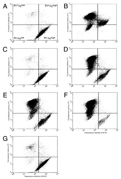
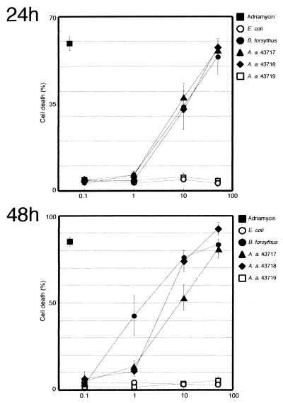
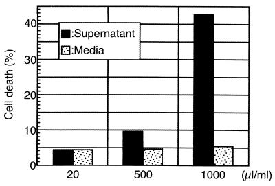
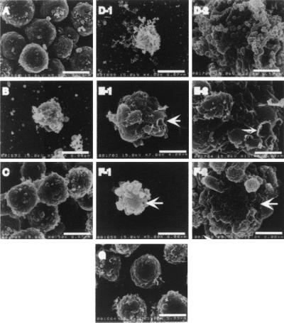
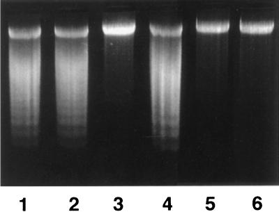
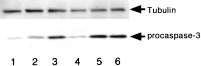
Bacteroides forsythus, which has been reported to be associated with periodontitis but has not been recognized as a key pathogen, was found to induce cytolytic activity against HL-60 and other human leukemic cells. This cytolytic activity was demonstrated according to three different criteria: (i) loss of both mitochondrial membrane potential and membrane integrity in cells treated with bacterial extracts and then with Rh123 and propidium iodide, respectively, as demonstrated by flow cytometry; (ii) damage to cytoplasmic membrane, as revealed by scanning electron microscopy (SEM); and (iii) DNA ladder formation and activation of caspase-3. These results indicate that B. forsythus produced an apoptosis-inducing factor(s) found to be composed of protein as judged by heat and trypsin sensitivity. In addition to extracts from B. forsythus, the culture supernatant of this bacterium has the ability to induce a cytolytic effect against peripheral white blood cells, especially lymphocytes. For comparison with B. forsythus, the same analyses were applied to two strains with different serotypes of Actinobacillus actinomycetemcomitans, serotypes a (ATCC 43717) and c (ATCC 43719), in addition to previously reported apoptosis-inducing serotype b (ATCC 43718), which was used as a positive control. The strains of A. actinomycetemcomitans serotypes a and b induced apoptosis in HL-60 cells as judged by the above three criteria but to a slightly lesser extent than did B. forsythus, while the serotype c strain produced apoptosis to a negligible extent. Detailed SEM images showed that the A. actinomycetemcomitans serotype a strain induced large-pore formation and the serotype b strain produced small pores with typical blebbing, while B. forsythus induced severe membrane ruffling. Further DNA ladder formation and caspase-3 activation were observed in the serotype a and b strains but not in the serotype c strain. The present paper is the first report of a protein factor(s) from B. forsythus and the A. actinomycetemcomitans serotype a strain which induces apoptotic cell death.


Figures

FIG. 1
FIG. 1
Flow cytometric analysis of HL-60 cells incubated with or without 50 μg of protein of the extracts from various bacteria per ml. Panels: A, PBS; B, adriamycin (10 μM); C, E. coli; D, B. forsythus; E, A. actinomycetemcomitans serotype a; F, A. actinomycetemcomitans serotype b; G, A. actinomycetemcomitans serotype c. The extent of cell death was assessed by measuring fluorescence intensity using a FACScalibur flow cytometer after staining with PI and Rh123. All analyses were performed in duplicate experiments.
FIG. 2
FIG. 2
Dose-dependent induction of apoptotic cell death by B. forsythus and A. actinomycetemcomitans strains. HL-60 cells were incubated in the presence of various concentrations of extracts, ranging from 0.1 to 50 μg of protein/ml for 24 or 48 h. All analyses were performed in duplicate experiments. The results shown are means and standard deviations.
FIG. 3
FIG. 3
Flow cytometric analysis of PWBC incubated with B. forsythus culture supernatant (20, 500, and 1,000 μl/ml) or corresponding volumes of uncultured medium. The extent of cell death caused by lymphocytes gated from PWBC was assessed by the method described in the legend to Fig. 1.
FIG. 4
FIG. 4
Ultrastructural changes of HL-60 cells incubated with or without 50 μg of protein of bacterial sonic extracts per ml. Panels: A, B, and C, control HL-60 cells (treated with PBS, 10 μM adriamycin, and E. coli extract, respectively); D, E, and F, cells incubated for 48 h in the presence of the extracts from B. forsythus, A. actinomycetemcomitans serotype a, and A. actinomycetemcomitans serotype b, respectively; D-2, E-2, and F-2, photographs of corresponding samples at a higher magnification; G, cells incubated in the presence of the extract from A. actinomycetemcomitans serotype c, exhibiting cell surfaces similar to those of untreated HL-60 cells. Arrows indicate pores on cell membranes. Bar lengths and magnifications: A, 5.96 μm, ×5,000; B, 6.00 μm, ×4,500; C, 6.67 μm, ×5,000; D-1, 6.67 μm, ×4,500; D-2, 1.50 μm, ×20,000; E-1, 4.29 μm, ×7,000; E-2, 2.00 μm, ×15,000; F-1, 6.00 μm, ×5,000; F-2, 2.00 μm, ×15,000; G, 5.99 μm, ×5,000. Overall shrinkage was observed in cells treated with B. forsythus and A. actinomycetemcomitans serotype a extracts, and the calculated diameters were 5.8 and 5.6 μm, respectively, while that of control cells was approximately 7.5 μm.
FIG. 5
FIG. 5
DNA ladder formation of HL-60 cells treated with bacterial extracts. HL-60 cells (5 × 10⁵) were treated with bacterial extracts (1 μg/ml) or PBS for 48 h at 37°C. Two micrograms of cellular DNA isolated using the Hirt method (7) was subjected to agarose gel electrophoresis and then stained with ethidium bromide. Lanes: 1, A. actinomycetemcomitans serotype a; 2, A. actinomycetemcomitans serotype b; 3, A. actinomycetemcomitans serotype c; 4, B. forsythus; 5, E. coli; 6, PBS. Nucleosomal DNA ladders characteristic of apoptotic cells were observed in the cells treated with B. forsythus, A. actinomycetemcomitans serotype a, and A. actinomycetemcomitans serotype b lysates.
FIG. 6
FIG. 6
Activation of caspase-3 by treatment of HL-60 cells in the presence of periodontopathic bacterial extracts. Ten micrograms of cellular lysate prepared from HL-60 cells (5 × 10⁵ cells) which had been treated with 1 μg of each bacterial extract per ml or with PBS was subjected to Western blot analysis by using anti-procaspase-3 antibody. Lanes: 1, A. actinomycetemcomitans serotype a; 2, A. actinomycetemcomitans serotype b; 3, A. actinomycetemcomitans serotype c; 4, B. forsythus; 5, E. coli; 6, PBS). Decreases in procaspase-3 levels represent the activation of caspase-3. The filter was also probed with anti-tubulin antibody to monitor the amount of loading in each lane.

References

    1. Arab S, Murakami M, Dirks P, Boyd B, Hubbard S L, Lingwood C A, Rutka J T. Verotoxins inhibit the growth of and induce apoptosis in human astrocytoma cells. J Neurooncol. 1998;40:137–150. - PubMed
    1. Bertrand S, Berger R, Philip T, Bernheim A, Bryon P A, Bertoglio J, Dore J F, Brunat-Mentigny M, Lenoir G M. Variant translocation in a non endemic case of Burkitt's lymphoma: t(8;22) in an Epstein-Barr virus negative tumor and in a derived cell line. Eur J Cancer. 1981;17:577–584. - PubMed
    1. Collins S J, Gallo R C, Gallagher R E. Continuous growth and differentiation of human myeloid leukemic cells in suspension culture. Nature. 1977;270:347–349. - PubMed
    1. Fives-Taylor P M, Meyer D H, Mintz K P, Brisette C. Virulence factors of Actinobacillus actinomycetemcomitans. Periodontol 2000. 1999;29:136–167. - PubMed
    1. Gewirtz D A. A critical evaluation of the mechanisms of action proposed for the antitumor effects of the anthracycline antibiotics adriamycin and daunorubicin. Biochem Pharmacol. 1999;57:727–741. - PubMed

Publication types

LinkOut - more resources