. 2000 Aug;68(8):4736-45.
doi: 10.1128/IAI.68.8.4736-4745.2000.

Nonopsonic phagocytosis of zymosan and Mycobacterium kansasii by CR3 (CD11b/CD18) involves distinct molecular determinants and is or is not coupled with NADPH oxidase activation

Affiliations

Nonopsonic phagocytosis of zymosan and Mycobacterium kansasii by CR3 (CD11b/CD18) involves distinct molecular determinants and is or is not coupled with NADPH oxidase activation

V Le Cabec et al. Infect Immun. 2000 Aug.

Abstract

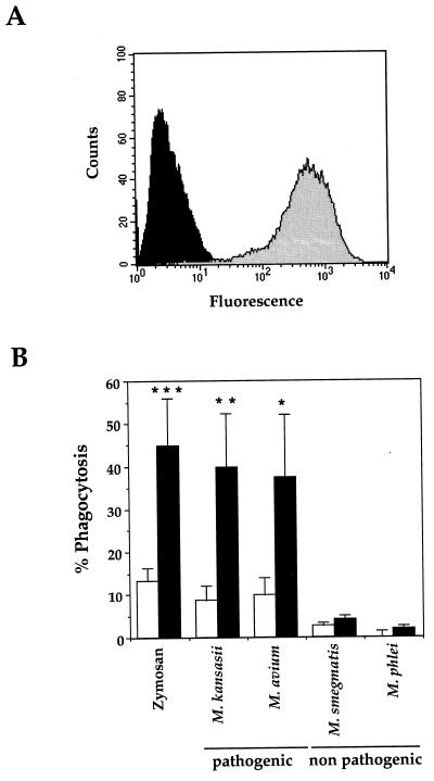
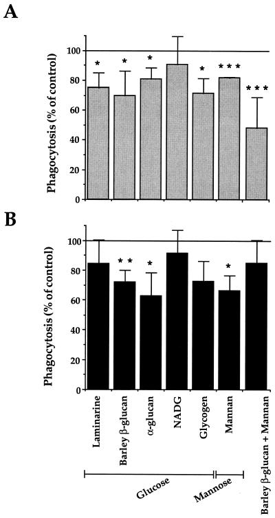
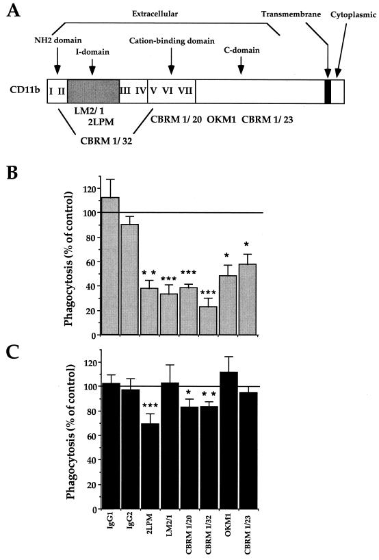
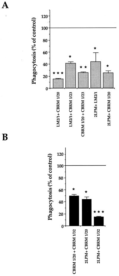
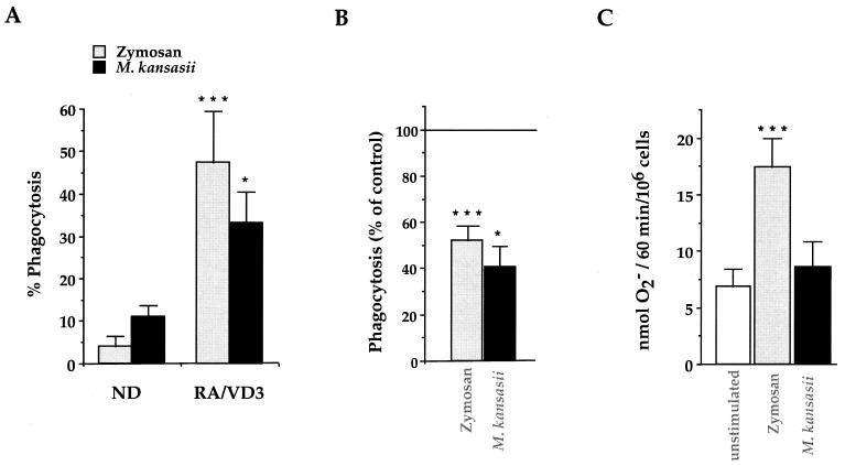
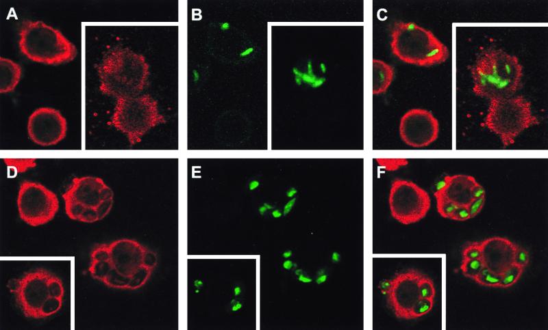
Complement receptor type 3 (CR3) was initially described as an opsonic receptor. Subsequently, CR3-mediated lectin-sugar recognition mechanisms have been shown to play a major role in the nonopsonic phagocytosis of several pathogens, among them Mycobacterium tuberculosis. Little is known about the binding and signal transduction mechanisms operating during nonopsonic ingestion through CR3 of different microorganisms. In the present study, we used CHO cells stably transfected with CR3 to show that CR3 was able to mediate internalization of zymosan and pathogenic mycobacteria (Mycobacterium kansasii and Mycobacterium avium) but not that of nonpathogenic species (Mycobacterium smegmatis and Mycobacterium phlei). A combination of mannan and beta-glucan inhibited the phagocytosis of zymosan but had no effect on M. kansasii ingestion. Among six monoclonal antibodies (MAbs) directed against the CD11b subunit of CR3 that decreased zymosan ingestion, only three inhibited M. kansasii phagocytosis. In particular, MAbs known to block the CR3 lectin site affected only internalization of zymosan. Using U937 macrophages, we observed that zymosan ingestion through CR3 induced superoxide production measured by cytochrome c reduction and by translocation of the NADPH oxidase cytosolic component p47phox to the phagosomal membrane, whereas phagocytosis of viable or heat-killed M. kansasii did not. Furthermore, lack of superoxide anion production during phagocytosis of M. kansasii was not due to inhibition of NADPH oxidase per se or superoxide anion scavenging. Together, our results indicate that (i) nonopsonic phagocytosis of zymosan and M. kansasii by CR3 implicates different molecular mechanisms involving multiple and distinct epitopes of CD11b and (ii) CR3 may transduce different cellular responses depending on the sites mediating nonopsonic phagocytosis.


Figures

FIG. 1
FIG. 1
Characterization of CR3-transfected CHO cells. (A) Surface expression of CR3 was analyzed by flow cytometry. Cells were stained with anti-CD11b MAbs (2LPM) and FITC-coupled rabbit anti-mouse secondary antibodies prior to fixation with paraformaldehyde (shaded histogram); in the control, the primary antibody was omitted (solid histogram). The histograms show the fluorescence (expressed in arbitrary units) measured on the complete cell population. (B) Phagocytosis of zymosan and the indicated species of pathogenic or nonpathogenic mycobacteria by WT (open bars) and CR3-expressing (solid bars) CHO cells was measured by fluorescence microscopy. At least 100 cells per slide were counted under fluorescence microscopy. The percentage of phagocytic cells (% Phagocytosis) having ingested at least one particle or bacterium (number of phagocytic cells/number of total cells × 100) is expressed as the means + SEM of 3 to 29 separate experiments performed in duplicate. Statistical differences were measured for CR3-transfected versus WT CHO cells. ∗, P < 0.05; ∗∗, P < 0.01; ∗∗∗, P < 0.005.
FIG. 2
FIG. 2
Inhibition by polysaccharides of CR3-mediated zymosan and M. kansasii phagocytosis. CR3-transfected CHO cells were preincubated as indicated with glucose- or mannose-containing polysaccharides or β-glucan plus mannan before incubation with either zymosan (A) or M. kansasii (B). The data are expressed as the percentage of phagocytosis reported compared to control values (100%, no polysaccharide). The values are means + SEM of three to four separate experiments performed in duplicate. Statistical differences were measured for cells pretreated with sugars compared to untreated cells. ∗, P < 0.05; ∗∗, P < 0.01; ∗∗∗, P < 0.005.
FIG. 3
FIG. 3
Inhibition of CR3-mediated phagocytosis of zymosan and M. kansasii by anti-CR3 MAbs. (A) Schematic map of the regions of the human CD11b α chain recognized by the different MAbs used in this study (3, 8, 31). (B and C) CR3-transfected CHO cells were preincubated with the indicated nonrelevant IgG1 or IgG2 or with anti-CD11b MAbs for 30 min at 37°C before incubation with zymosan (B) or M. kansasii (C). The data are expressed as the percentage of phagocytosis compared to control values (100%, no antibody). The values are means + SEM of three to six separate experiments performed in duplicate. Statistical differences were measured for cells pretreated with MAbs compared to untreated cells. ∗, P < 0.05; ∗∗, P < 0.01; ∗∗∗, P < 0.005.
FIG. 4
FIG. 4
Inhibition of the phagocytosis of zymosan and M. kansasii by CR3-transfected CHO cells by anti-CR3 MAb combinations. As described in the legend to Fig. 3, the cells were preincubated with the indicated combinations of MAbs before incubation with zymosan (A) or M. kansasii (B). The values are the means + SEM of three separate experiments performed in duplicate. Statistical differences were measured for cells pretreated with MAbs compared to untreated cells. ∗, P < 0.05; ∗∗, P < 0.01; ∗∗∗, P < 0.005.
FIG. 5
FIG. 5
Bactericidal responses of RA-VD3-differentiated U937 cells in response to zymosan and M. kansasii. (A) Phagocytosis of zymosan and M. kansasii was measured in undifferentiated (ND) and differentiated (RA/VD3) U937 cells after 1 h of incubation. The data are expressed as the percentage of phagocytosis, and the values are the means + SEM of three to six separate experiments performed in duplicate. Statistical differences were measured for differentiated cells compared to undifferentiated cells. (B) RA-VD3-differentiated U937 cells were preincubated with 2LPM MAbs prior to incubation with zymosan or M. kansasii to assess the involvement of CR3 in particle internalization. The cells were then processed for measurement of phagocytosis. The data are expressed as the percentage of phagocytosis reported compared to control values, and the values are the means + SEM of three to six separate experiments performed in duplicate. Statistical differences were calculated for cells pretreated with MAbs compared to untreated cells. (C) RA-VD3-differentiated U937 cells were incubated for 1 h either alone (open bar) or in the presence of zymosan or M. kansasii and processed for measurement of O2 production. The data are expressed in nanomoles of O2 produced by 106 cells in 1 h, and the values are the means + SEM of three separate experiments performed in duplicate. Statistical differences were measured for cells incubated with zymosan or M. kansasii compared to unstimulated cells. ∗, P < 0.05; ∗∗∗, P < 0.005.
FIG. 6
FIG. 6
Analysis of NADPH oxidase assembly at the phagosomal membrane by confocal microscopy. Phagocytosis of FITC-labeled M. kansasii (A to C) or FITC-labeled zymosan (D to F) was not synchronized in order to obtain phagosomes at different maturation stages. Following phagocytosis, differentiated U937 cells were fixed, permeabilized, and stained with anti-p47phox antibodies revealed by TRITC-conjugated anti-rabbit antibodies. The stained cells are representative of three independent experiments, and phagosomes visualized by confocal microscopy are shown. (A and D) p47phox staining alone; (B and E) FITC-labeled particles alone; (C and F) superimposition of both types of staining. Insets, representative of a second experiment.

References

    1. Armstrong J A, Hart P D. Phagosome-lysosome interactions in cultured macrophages infected with virulent tubercle bacilli. J Exp Med. 1975;142:1–16. - PMC - PubMed
    1. Astarie-Dequeker C, N'Diaye E N, Le Cabec V, Rittig M G, Prandy J, Maridonneau-Parini I. The mannose receptor mediates uptake of pathogenic and nonpathogenic mycobacteria and bypasses bactericidal responses in human macrophages. Infect Immun. 1999;67:469–477. - PMC - PubMed
    1. Balsam L B, Liang T W, Parkos C A. Functional mapping of CD11b/CD18 epitopes important in neutrophil-epithelial interactions: a central role of the I domain. J Immunol. 1998;160:5058–5065. - PubMed
    1. Bermudez L E, Goodman J, Petrofsky M. Role of complement receptors in uptake of Mycobacterium avium by macrophages in vivo: evidence from studies using CD18-deficient mice. Infect Immun. 1999;67:4912–4916. - PMC - PubMed
    1. Bermudez L E, Parker A, Goodman J R. Growth within macrophages increases the efficiency of Mycobacterium avium in invading other macrophages by a complement receptor-independent pathway. Infect Immun. 1997;65:1916–1925. - PMC - PubMed

Publication types

LinkOut - more resources