. 2000 Jul 17;192(2):159-70.
doi: 10.1084/jem.192.2.159.

Metabolic consequences of adenosine deaminase deficiency in mice are associated with defects in alveogenesis, pulmonary inflammation, and airway obstruction

Affiliations

Metabolic consequences of adenosine deaminase deficiency in mice are associated with defects in alveogenesis, pulmonary inflammation, and airway obstruction

M R Blackburn et al. J Exp Med. .

Abstract

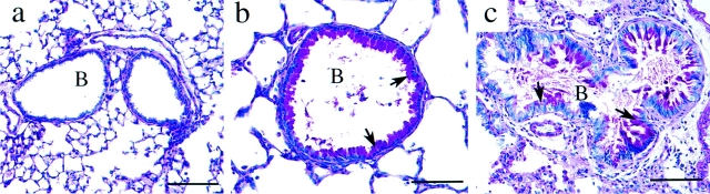
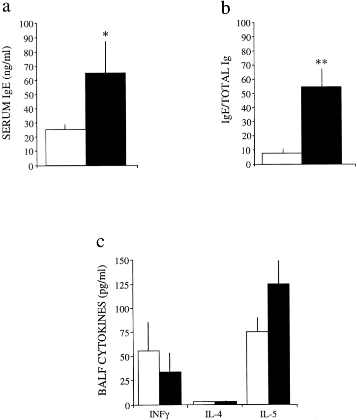
Adenosine deaminase (ADA) is a purine catabolic enzyme that manages levels of the biologically active purines adenosine and 2'-deoxyadenosine in tissues and cells. ADA-deficient mice die at 3 wk of age from severe respiratory distress. This phenotype is progressive and is linked to perturbations in pulmonary purine metabolism. The inflammatory changes found in the lungs of ADA-deficient mice included an accumulation of activated alveolar macrophages and eosinophils. These changes were accompanied by a pronounced enlargement of alveolar spaces and increases in mucus production in the bronchial airways. The alveolar enlargement was found to be due in part to abnormal alveogenesis. Lowering adenosine and 2'-deoxyadenosine levels using ADA enzyme therapy decreased the pulmonary eosinophilia and resolved many of the lung histopathologies. In addition, genetically restoring ADA to the forestomach of otherwise ADA-deficient mice prevented adenine metabolic disturbances as well as lung inflammation and damage. These data suggest that disturbances in purinergic signaling mediate the lung inflammation and damage seen in ADA-deficient mice.


Figures

Figure 1
Figure 1
Morphological and cytological changes in the lungs of ADA-deficient mice. (a) H&E-stained control lung at postpartum day 18. (b) H&E-stained ADA-deficient lung at postpartum day 18. Arrows indicate areas of inflammation. Notice the pronounced enlargement of alveolar spaces (AS) and the thickening of pulmonary blood vessels (BV). (c) High magnification of an H&E-stained ADA-deficient lung demonstrating eosinophil infiltration (arrows) around bronchioles (B) and pulmonary blood vessels (BV). (d) Immunolocalization of eosinophils in an 18-d-old ADA-deficient lung, using a rabbit polyclonal antibody raised against mMBP-1 followed by detection with FITC immunofluorescence. Notice intense staining of inflammatory cells with anti–MBP-1 (arrows). mMBP-1 immunoreactivity was also detected in alveolar macrophages (AM). Cytological analysis of cells collected from BALF from the lung of an 18-d-old ADA-deficient mouse showing (e) eosinophils (arrows), (f) alveolar macrophages engulfing eosinophils (arrows), and (g) multinucleated giant cells. Bars, (a–d) 100 μm; (e–g) 10 μm.
Figure 2
Figure 2
Mucus hypersecretion in the bronchial airways of ADA-deficient mice. Lung sections were stained with PAS for the detection of neutral mucins. (a) Control lung at postpartum day 18. (b) ADA-deficient lung at postpartum day 18. (c) ADA-deficient lung at postnatal day 21. Arrows in b and c denote PAS-positive material indicative of increased mucus production. B, bronchiole. Bars, 100 μm.
Figure 3
Figure 3
Defects in alveogenesis in ADA-deficient mice. Lungs from age-matched control and ADA-deficient mice were collected and processed for H&E staining. (a) Control lung at postpartum day 0. (b) ADA-deficient lung at day 0. (c) Control lung at day 5. (d) ADA-deficient lung at day 5. (e) Control lung at day 10 demonstrating the septation of presumptive alveoli into mature alveolar sacs. (f) ADA-deficient lung at day 10 demonstrating enlarged alveolar spaces. Panels a–f are at the same magnification; bars, 250 μm. (g) The size of alveolar airways was determined in control (white bars, n = 4) and ADA-deficient (black bars, n = 4) lungs by measuring mean chord lengths (in μm) of alveolar airways in H&E-stained lungs. Values are given as mean μm ± SE from four separate age-matched control and ADA-deficient lung pairs at each developmental stage; *P ≤ 0.05.
Figure 3
Figure 3
Defects in alveogenesis in ADA-deficient mice. Lungs from age-matched control and ADA-deficient mice were collected and processed for H&E staining. (a) Control lung at postpartum day 0. (b) ADA-deficient lung at day 0. (c) Control lung at day 5. (d) ADA-deficient lung at day 5. (e) Control lung at day 10 demonstrating the septation of presumptive alveoli into mature alveolar sacs. (f) ADA-deficient lung at day 10 demonstrating enlarged alveolar spaces. Panels a–f are at the same magnification; bars, 250 μm. (g) The size of alveolar airways was determined in control (white bars, n = 4) and ADA-deficient (black bars, n = 4) lungs by measuring mean chord lengths (in μm) of alveolar airways in H&E-stained lungs. Values are given as mean μm ± SE from four separate age-matched control and ADA-deficient lung pairs at each developmental stage; *P ≤ 0.05.
Figure 4
Figure 4
Levels of serum IgE and BALF cytokines in control (white bars) and ADA-deficient (black bars) mice. (a) Total IgE levels were measured in the serum of 18-d-old control (n = 11) and ADA-deficient (n = 8) mice. Mean total serum IgE values are given as ng/ml ± SE. Total serum Ig [IgG + IgM] levels are decreased 67% in ADA-deficient mice (reference 15). (b) Ratios of IgE to Ig in control and ADA-deficient mice, demonstrating a significant bias towards hyper IgE in ADA-deficient mice. Statistical significance was determined using Student's t test analysis; *P ≤ 0.100, **P ≤ 0.05. (c) The levels of IFN-γ, IL-4, and IL-5 were measured in BALF collected from 18-d-old control (n = 10) and ADA-deficient (n = 7) mice. Mean values are given as pg/ml ± SE.
Figure 6
Figure 6
Quantification of lung histopathology in ADA-deficient mice and ADA-deficient mice treated with PEG-ADA. (a) Mean cord lengths of alveolar airways were determined in lungs of 18-d-old control and ADA-deficient mice, and lungs of 21-d-old mice 72 h after treatment with PEG-ADA. Values are given as mean cord lengths in μm ± SE, n = 5 for each condition. *P ≤ 0.02 for increases in mean cord lengths compared with controls. (b) The degree of mucus production was determined in the bronchial airways of 18-d-old control and ADA-deficient mice, and 21-d-old ADA-deficient mice 3 d after treatment with PEG-ADA. Values are given as mean airway mucus indices in arbitrary pixel units ± SE, n = 5 for each condition. Statistical significance was determined using Student's t test analysis; *P ≤ 0.01 for an increase in mucus index between control and ADA-deficient samples, **P ≤ 0.02 for a decrease in mucus index between ADA-deficient samples and PEG-ADA–treated ADA-deficient samples.
Figure 5
Figure 5
Reversible lung eosinophilia in ADA-deficient mice treated with PEG-ADA. Total cellular differentials were determined on cells collected from BALF of 18-d-old control mice (n = 19, white bars), ADA-deficient mice (n = 12, black bars), and ADA-deficient mice treated with PEG-ADA and examined 3 d later (n = 5, hatched bars) or after 2 wk of enzyme therapy (n = 4, stippled bars). Mean values are given as total cells ± SE. Statistical significance was determined using Student's t test analysis; *P ≤ 0.02.
Figure 7
Figure 7
PEG-ADA treatments reverse accumulations of adenosine and 2′-deoxyadenosine in the lungs of ADA-deficient mice. Adenosine and 2′-deoxyadenosine levels were quantitated in the lungs of 18-d-old control mice (n = 5) and ADA-deficient mice (n = 4), and 21-d-old ADA-deficient mice 3 d after treatment with PEG-ADA (n = 4). Mean values are given as nmol/mg protein ± SE. Statistical significance was determined using Student's t test analysis; *P ≤ 0.002. nd, not detectable at a minimal detection limit of 0.001 nmol/mg protein.
Figure 8
Figure 8
Transgenic expression of ADA in the forestomach of ADA-deficient mice prevents adenosine and 2′-deoxyadenosine accumulation, lung inflammation, and lung histopathologies. (a) H&E-stained section of an 18-d-old control lung. (b) H&E-stained section of a 21-d-old ADA-deficient lung. (c) H&E-stained section of a 21-d-old ADA-deficient lung of a mouse expressing an ADA minigene in its forestomach. Bars, (a–c) 250 μm. (d) Adenosine and 2′-deoxyadenosine levels were quantitated in the lungs of 21-d-old control, ADA-deficient, or ADA-deficient mice expressing ADA in their forestomach (forestomach rescue). Mean values are given as nmol/mg protein ± SE; n = 3 for each. Statistical significance was determined using Student's t test analysis; *P ≤ 0.002. nd, not detected at a minimal detection limit of 0.001 nmol/mg protein. (e) Zymogram analysis showing the level of ADA enzymatic activity in the lung (L), blood (Bl), and forestomach (FS) of 21-d-old control, ADA-deficient, and ADA-deficient mice expressing ADA in their forestomach. Purine nucleoside phosphorylase (PNP) was used as a positive control.

References

    1. Frederiksen S. Specificity of adenosine deaminase toward adenosine and 2′-deoxyadenosine analogues. Arch. Biochem. Biophys. 1966;113:383–388. - PubMed
    1. Olah M.E., Stiles G.L. Adenosine receptor subtypescharacterization and therapeutic regulation. Annu. Rev. Pharmacol. Toxicol. 1995;35:581–606. - PubMed
    1. Hershfield M.S., Kredich N.M., Ownby D.R., Ownby H., Buckley R. In vivo inactivation of erythrocyte S-adenosylhomocysteine hydrolase by 2′-deoxyadenosine in adenosine deaminase–deficient patients. J. Clin. Invest. 1979;63:807–811. - PMC - PubMed
    1. Benveniste P., Zhu W., Cohen A. Interference with thymocyte differentiation by an inhibitor of S-adenosylhomocysteine hydrolase. J. Immunol. 1995;155:536–544. - PubMed
    1. Benveniste P., Cohen A. p53 expression is required for thymocyte apoptosis induced by adenosine deaminase deficiency. Proc. Natl. Acad. Sci. USA. 1995;92:8373–8377. - PMC - PubMed

Publication types