. 2000 Jul 17;192(2):303-10.
doi: 10.1084/jem.192.2.303.

Immunologic self-tolerance maintained by CD25(+)CD4(+) regulatory T cells constitutively expressing cytotoxic T lymphocyte-associated antigen 4

Affiliations

Immunologic self-tolerance maintained by CD25(+)CD4(+) regulatory T cells constitutively expressing cytotoxic T lymphocyte-associated antigen 4

T Takahashi et al. J Exp Med. .

Abstract

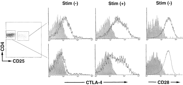
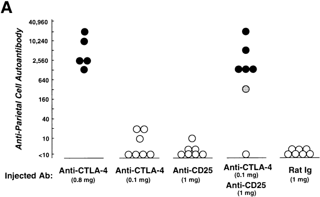
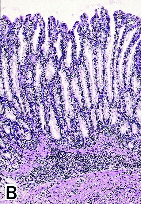
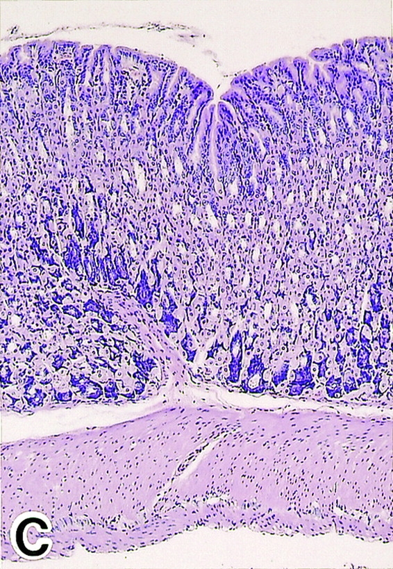
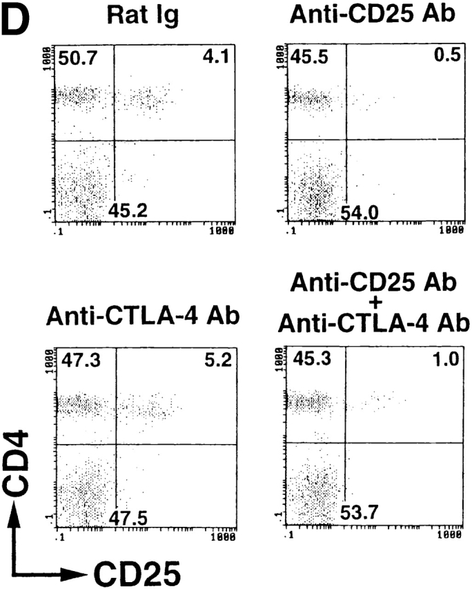
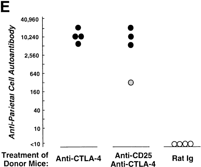
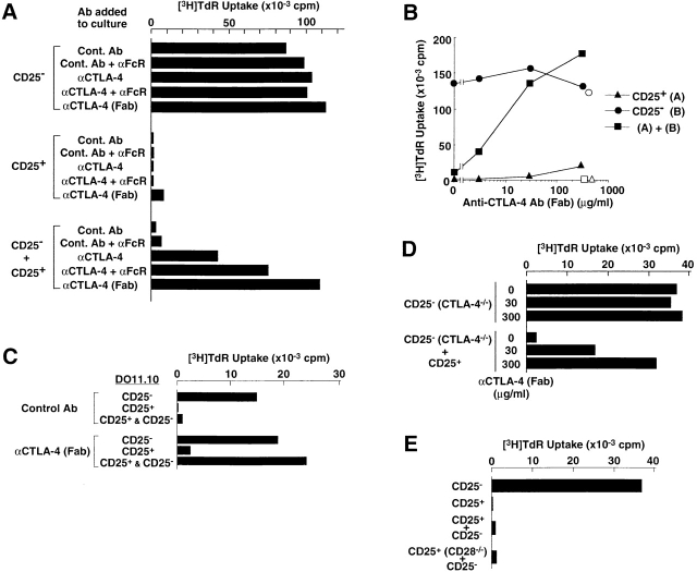
This report shows that cytotoxic T lymphocyte-associated antigen 4 (CTLA-4) plays a key role in T cell-mediated dominant immunologic self-tolerance. In vivo blockade of CTLA-4 for a limited period in normal mice leads to spontaneous development of chronic organ-specific autoimmune diseases, which are immunopathologically similar to human counterparts. In normal naive mice, CTLA-4 is constitutively expressed on CD25(+)CD4(+) T cells, which constitute 5-10% of peripheral CD4(+) T cells. When the CD25(+)CD4(+) T cells are stimulated via the T cell receptor in vitro, they potently suppress antigen-specific and polyclonal activation and proliferation of other T cells, including CTLA-4-deficient T cells, and blockade of CTLA-4 abrogates the suppression. CD28-deficient CD25(+)CD4(+) T cells can also suppress normal T cells, indicating that CD28 is dispensable for activation of the regulatory T cells. Thus, the CD25(+)CD4(+) regulatory T cell population engaged in dominant self-tolerance may require CTLA-4 but not CD28 as a costimulatory molecule for its functional activation. Furthermore, interference with this role of CTLA-4 suffices to elicit autoimmune disease in otherwise normal animals, presumably through affecting CD25(+)CD4(+) T cell-mediated control of self-reactive T cells. This unique function of CTLA-4 could be exploited to potentiate T cell-mediated immunoregulation, and thereby to induce immunologic tolerance or to control autoimmunity.


Figures

Figure 1
Figure 1
Constitutive expression of CTLA-4 on CD25+CD4+ T cells in normal naive mice. Lymph node and spleen cell suspensions prepared from a 2-mo-old BALB/c mouse were stained with FITC–anti-CD4 Ab and biotinylated anti-CD25 Ab with RPE-CyChrome 5–conjugated streptavidin as the secondary reagent. Cells enclosed with rectangles were sorted as CD25+ or CD254+ T cells. The sorted cells were stained for 2 h at 37°C with PE-conjugated anti–CTLA-4 Ab or PE-conjugated anti–human CD4 Ab as a staining control, and are shown as histograms (solid lines or shaded areas, respectively; the abscissa shows the staining intensity on a logarithmic scale and the ordinate shows the number of cells on an arbitrary scale) (reference 28). The sorted cells were also cultured with Con A (1 μg/ml) at 37°C for 24 h and incubated for the last 2 h with PE–anti–CTLA-4 Ab. CD28 expression on CD25+ or CD254+ T cells is shown as histograms by staining the cells with PE–anti-CD28 Ab. A representative result of five independent experiments is shown. Stim., stimulation.
Figure 2
Figure 2
Induction of autoimmune disease in BALB/c mice by administration of anti–CTLA-4 Ab with or without anti-CD25 Ab. (A) BALB/c mice were injected intraperitoneally with indicated doses of purified anti–CTLA-4 mAb, anti-CD25 mAb, a mixture of the two mAbs, or control rat Ig once a week three times from the age of 10 d, and were examined at 3 mo of age for histological and/or serological development of autoimmune disease. The gastric lesion was histologically graded as macroscopically and histologically evident grade-2 gastritis (black circle), histologically evident grade-1 gastritis (gray circle), or intact gastric mucosa (white circle; for detailed grading, see references 12, 14, 15). Titers of antiparietal cell autoantibodies were assessed by ELISA. The gastric mucosa of a grade-2 gastritis in a mouse treated with anti–CTLA-4 mAb is shown in B, and the intact gastric mucosa of a mouse treated with rat Ig is shown in C. Samples were stained with hematoxylin and eosin (original magnification: ×25). (D) Composition of T cell subpopulations defined by the expression of CD4 and CD25 7 d after the last Ab treatment. Lymph node cells were stained with PE–anti-CD4 mAb and FITC–anti-CD25 mAb, 7D4, another anti-CD25 mAb with different epitope specificity, as described previously (reference 18). A representative staining of three independent experiments is shown. (E) Spleen cells (5 × 10⁷) from anti–CTLA-4 mAb– or mixed mAb–treated mice with gastritis or from rat Ig–treated mice were transferred intravenously to BALB/c athymic nude mice, which were histologically and serologically examined 2 mo later. Black circle, macroscopically and histologically evident grade-2 gastritis; gray circle, histologically evident grade-1 gastritis; white circle, intact gastric mucosa. A representative result of more than three independent experiments is shown. The means of duplicate cultures are shown, and the SEM was within 10% of the mean for each experiment.
Figure 2
Figure 2
Induction of autoimmune disease in BALB/c mice by administration of anti–CTLA-4 Ab with or without anti-CD25 Ab. (A) BALB/c mice were injected intraperitoneally with indicated doses of purified anti–CTLA-4 mAb, anti-CD25 mAb, a mixture of the two mAbs, or control rat Ig once a week three times from the age of 10 d, and were examined at 3 mo of age for histological and/or serological development of autoimmune disease. The gastric lesion was histologically graded as macroscopically and histologically evident grade-2 gastritis (black circle), histologically evident grade-1 gastritis (gray circle), or intact gastric mucosa (white circle; for detailed grading, see references 12, 14, 15). Titers of antiparietal cell autoantibodies were assessed by ELISA. The gastric mucosa of a grade-2 gastritis in a mouse treated with anti–CTLA-4 mAb is shown in B, and the intact gastric mucosa of a mouse treated with rat Ig is shown in C. Samples were stained with hematoxylin and eosin (original magnification: ×25). (D) Composition of T cell subpopulations defined by the expression of CD4 and CD25 7 d after the last Ab treatment. Lymph node cells were stained with PE–anti-CD4 mAb and FITC–anti-CD25 mAb, 7D4, another anti-CD25 mAb with different epitope specificity, as described previously (reference 18). A representative staining of three independent experiments is shown. (E) Spleen cells (5 × 10⁷) from anti–CTLA-4 mAb– or mixed mAb–treated mice with gastritis or from rat Ig–treated mice were transferred intravenously to BALB/c athymic nude mice, which were histologically and serologically examined 2 mo later. Black circle, macroscopically and histologically evident grade-2 gastritis; gray circle, histologically evident grade-1 gastritis; white circle, intact gastric mucosa. A representative result of more than three independent experiments is shown. The means of duplicate cultures are shown, and the SEM was within 10% of the mean for each experiment.
Figure 3
Figure 3
Key role of CTLA-4 expressed on CD25+CD4+ regulatory T cells in downregulating the activation and proliferation of other T cells. (A) Anti–CTLA-4 mAb (100 μg/ml) or control hamster Ab with or without anti-FcR mAb (10 μg/ml) or Fab–anti-CTLA-4 mAb (100 μg/ml) was added to the culture of CD25+ or CD25CD4+ T cells purified from BALB/c spleen and lymph node cells (as shown in Fig. 1), or these two populations were mixed at a 1:1 ratio. The culture was stimulated for 3 d with Con A (1 μg/ml) along with irradiated BALB/c spleen cells as APCs. Incorporation of 3H-TdR during the last 6 h of the culture is shown as the degree of proliferation. Background counts in the wells with APCs alone were always <1,500 cpm in these experiments and those below. α, anti-. (B) CD25+ or CD25CD4+ T cells or a 1:1 mixture of the two populations were stimulated with anti-CD3 mAb (10 μg/ml) in the presence of Fab–anti–CTLA-4 mAb at various concentrations (black symbols) or control Fab–hamster IgG at 300 μg/ml (white symbols). (C) CD25+ or CD25CD4+ T cells or two populations mixed at an equal ratio, purified from the spleens of DO11.10 transgenic mice, were cultured for 3 d with 0.1 μM OVA-P(323–339) in the presence of Fab–anti–CTLA-4 mAb or control Fab–hamster IgG at 100 μg/ml. α, anti-. (D) CD25CD4+ T cells prepared from 3-wk-old CTLA-4–deficient (CTLA-4−/−) B6 mice or a 1:1 mixture of these cells with CD25+CD4+ T cells from 2-mo-old normal B6 mice was stimulated with anti-CD3 mAb for 3 d in the presence of Fab–anti–CTLA-4 mAb at indicated concentrations. The counts of CTLA-4–deficient CD25CD4+ T cells cultured without stimulation were background levels (i.e., <2,000 cpm). α, anti-. (E) CD25+CD4+ T cells prepared from CD28-deficient (CD28−/−) B6 mice or normal B6 mice were mixed with an equal number of CD25CD4+ T cells from normal B6 mice and stimulated with anti-CD3 mAb for 3 d. A representative result of more than three independent experiments is shown. The means of duplicate cultures are shown, and the SEM was always with 10% of the mean for each experiment.

References

    1. Thompson C.B., Allison J.P. The emerging role of CTLA-4 as an immune attenuator. Immunity. 1997;7:445–450. - PubMed
    1. Bluestone J.A. Is CTLA-4 a master switch for peripheral T cell tolerance? J. Immunol. 1997;158:1989–1993. - PubMed
    1. Waterhouse P., Penninger J.M., Timms E., Wakeham A., Shahinian A., Lee K.P., Thompson C.B., Griesser H., Mak T.W. Lymphoproliferative disorders with early lethality in mice deficient in Ctla-4. Science. 1995;270:985–988. - PubMed
    1. Tivol E.A., Borriello F., Schweitzer A.N., Lynch W.P., Bluestone J.A., Sharpe A.H. Loss of CTLA-4 leads to massive lymphoproliferation and fatal multiorgan tissue destruction, revealing a critical negative regulatory role of CTLA-4. Immunity. 1995;3:541–547. - PubMed
    1. Karandikar N.J., Vanderlugt C.L., Walunas T.L., Miller S.D., Bluestone J.A. CTLA-4a negative regulator of autoimmune disease. J. Exp. Med. 1996;184:783–788. - PMC - PubMed

Publication types