. 2000 Jul;12(7):1179-201.
doi: 10.1105/tpc.12.7.1179.

The C-terminal dilysine motif confers endoplasmic reticulum localization to type I membrane proteins in plants

Affiliations

The C-terminal dilysine motif confers endoplasmic reticulum localization to type I membrane proteins in plants

M Benghezal et al. Plant Cell. 2000 Jul.

Abstract

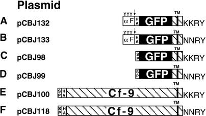
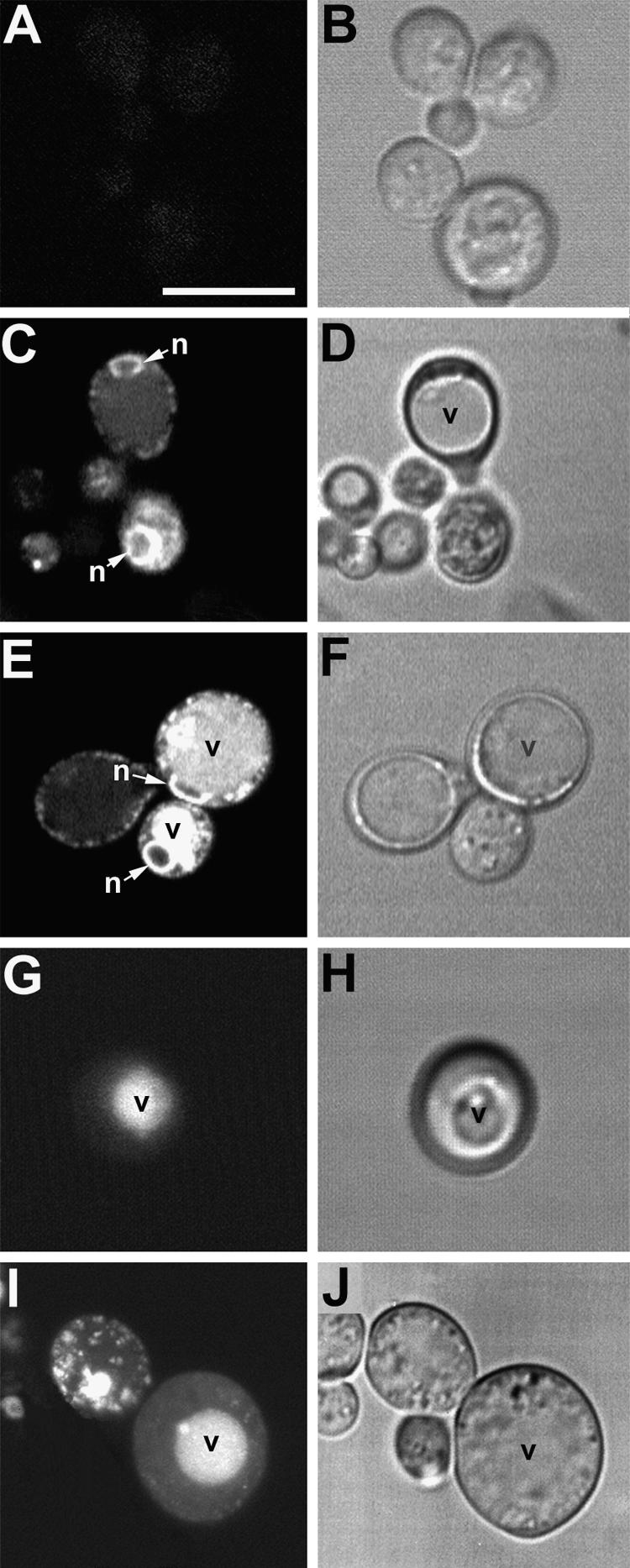
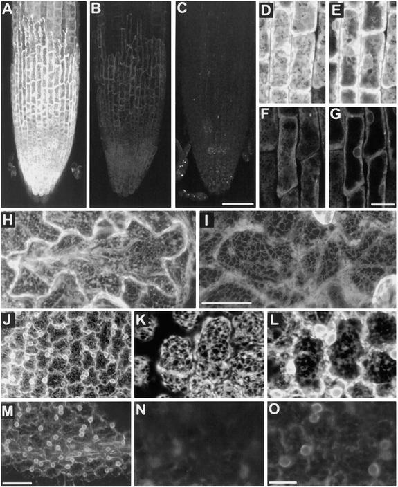
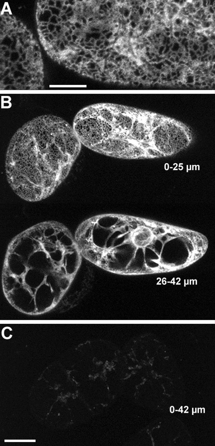
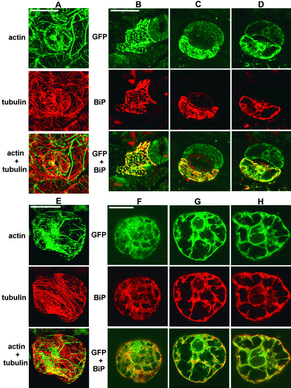
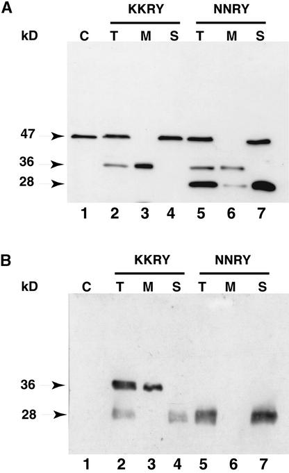
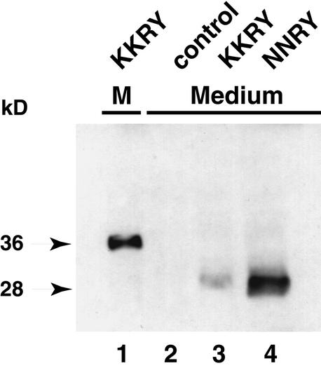
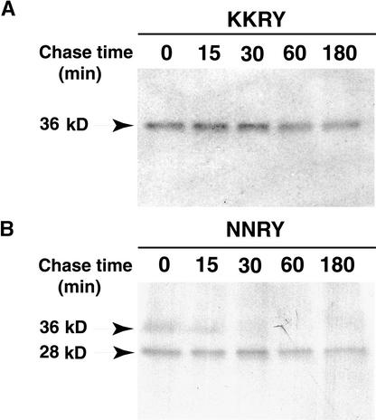
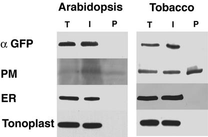
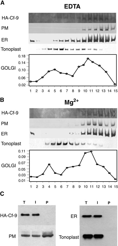
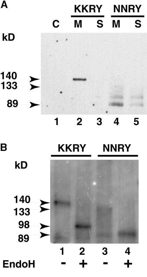
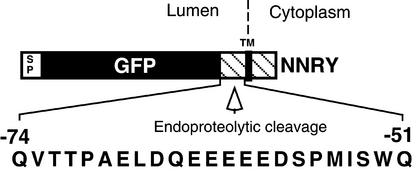
The tomato Cf-9 disease resistance gene encodes a type I membrane protein carrying a cytosolic dilysine motif. In mammals and yeast, this motif promotes the retrieval of type I membrane proteins from the Golgi apparatus to the endoplasmic reticulum (ER). To test whether the C-terminal KKXX signal of Cf-9 is functional as a retrieval motif and to investigate its role in plants, green fluorescent protein (GFP) was fused to the transmembrane domain of Cf-9 and expressed in yeast, Arabidopsis, and tobacco cells. The fusion protein was targeted to the ER in each of these expression systems, and mutation of the KKXX motif to NNXX led to secretion of the fusion protein. In yeast, the mutant protein reached the vacuole, but plants secreted it as a soluble protein after proteolytic removal of the transmembrane domain. Triple hemagglutinin (HA)-tagged full-length Cf-9 was also targeted to the ER in tobacco cells, and cleavage was also observed for the NNXX mutant protein, suggesting an endoprotease recognition site located within the Cf-9 lumenal sequence common to both the GFP- and the HA-tagged fusions. Our results indicate that the KKXX motif confers ER localization in plants as well as mammals and yeast and that Cf-9 is a resident protein of the ER.


Figures

Figure 1.
Figure 1.
Fusion Protein Constructs Analyzed in This Study and Their Corresponding Plasmids. (A) and (B) Constructs designed for expression in yeast, under the copper-inducible promoter. The first 85 amino acids of the yeast prepro-α factor (αF), containing a signal peptide and three N-glycosylation sites (Y), allow correct ER translocation. The triple HA epitope tag is indicated, and GFP is highlighted in black. The Cf-9 sequence is hatched, and the position of the transmembrane (TM) domain is also indicated. The arrow shows the position of the cleavage site for the late Golgi Kex2 endopeptidase. (C) to (F) Constructs designed for expression in Arabidopsis and tobacco BY-2 cells, under the cauliflower mosaic virus (CaMV) 35S promoter. The Arabidopsis chitinase signal peptide (SP) confers efficient ER translocation. Constructs in (A) to (D) carry only domains E (acidic), F (transmembrane), and G (basic, cytosolic) of Cf-9 (Jones et al., 1994), whereas constructs in (E) and (F) also carry domains B, C, and D of Cf-9 (Jones et al., 1994). The four amino acids corresponding to the Cf-9 C-terminal dilysine motif or its mutant derivative are shown at right.
Figure 2.
Figure 2.
Distribution of GFP Fluorescence in Yeast Demonstrates That the Cf-9 Dilysine Motif Confers ER Localization to Membrane-Bound GFP. (A) and (B) Nontransformed yeast cells are only weakly autofluorescent (∼1-μm section through midplanes of cells). (C) and (D) Localization pattern of the KKRY fusion. A midplane section (∼1 μm) reveals a perinuclear (n) and punctate peripheral fluorescence pattern, consistent with ER localization. Note the absence of fluorescence in the vacuole (v), which is clearly visible in the bright- field image (D). (E) and (F) Overexpression of the KKRY fusion results in secretion of the fusion protein into the vacuoles (v) of some cells. n, nucleus. (G) and (H) Localization of the NNRY fusion. A projection of seven sequential scans through the entire cell (total depth, ∼6 μm) demonstrates a strictly vacuolar fluorescence pattern (v) with no ER distribution pattern. (I) and (J) Overexpression of the NNRY fusion results in a punctate fluorescence in addition to the vacuolar localization pattern (v), consistent with Golgi localization. The confocal projection in (I) includes 10 consecutive sections (total depth, ∼8.5 μm). Confocal fluorescence ([A], [C], [E], [G], and [I]) and bright-field (transmission mode) images ([B], [D], [F], [H], and [J]) were obtained using identical acquisition and image processing parameters. (G) and (I) are projections of several optical sections, so their background areas appear relatively bright. formula image.
Figure 3.
Figure 3.
The Cf-9 Dilysine Motif Prevents Processing of Membrane-Bound GFP in Yeast. Microsomal proteins of transformed BJ2168 yeast cells were analyzed by SDS-PAGE on a 12% gel, processed for protein gel blotting, and probed with anti-GFP-specific antibody. Lane 1 shows proteins from yeast transformed with YEplac195 (vector only); lane 2, proteins from yeast transformed with pCBJ132 (KKRY fusion); and lane 3, proteins from yeast transformed with pCBJ133 (NNRY fusion). The relative molecular masses of the protein bands are indicated at left in kilodaltons.
Figure 4.
Figure 4.
Differences in GFP Fluorescence in the ER of Arabidopsis Demonstrate That the Cf-9 Dilysine Motif Confers ER Localization to Membrane-Bound GFP. Relative fluorescence of a KKRY transformant ([A], [D], [E], [H], and [J] to [L]) compared with a NNRY transformant ([B], [F], [G], [I], and [M] to [O]) in three different tissues. (A) to (C) Root cap epidermal cells. At identical acquisition settings, the intensity of fluorescence is markedly greater in the KKRY transformant (A), despite greater expression levels of the NNRY fusion (B). Autofluorescence of a nontransformed plant (C) is distinct from that of the GFP fusion products, demonstrating that the fluorescence in (A) and (B) is GFP specific. (D) and (E) Root cap epidermis of KKRY fusion. (D) shows an outer surface (∼6 μm thick) projection and (E) a midplane (∼3 μm) projection. (F) and (G) Root cap epidermis of NNRY fusion. (F) shows an outer surface (∼6 μm thick) projection and (G) a midplane (∼3 μm) projection. Note cortical ER ([D] and [F]) and perinuclear ([E] and [G]) localization. (H) and (I) Comparison of KKRY (H) and NNRY (I) fusion protein fluorescence in cotyledon pavement cells, abaxial surface. Note the weaker fluorescence in (I) and the strong delineation of the anticlinal cell surface in (H), despite identical thickness (19 μm) Z-series projections. (J) to (O) Comparison of KKRY ([J] to [L]) and NNRY ([M] to [O]) fusion fluorescence in petal epidermal cells. (J) KKRY fusion with projection of 27 1-μm-thick sections. (K) and (L) KKRY fusion, with higher magnification image of petal epidermis, with 7.2-μm projection through cell periphery showing cortical ER network (K) and 10.4-μm projection through cell midplane showing strong perinuclear fluorescence (L). (M) NNRY fusion, with as in (J), a projection of 27 1-μm-thick optical sections. (N) and (O) NNRY fusion. (N) Higher magnification image of petal epidermis; 7.2-μm projection through cell periphery. Compared with (K), there is almost no fluorescence in the cortical ER. (O) Midplane projection (10.4 μm thick) immediately beneath the projection shown in (N). Some perinuclear fluorescence is detected. formula image; formula image; formula image; formula image; formula image.
Figure 5.
Figure 5.
The Dilysine Motif Confers ER Localization to Membrane-Bound GFP in Tobacco BY-2 Cells. (A) KKRY fusion. A projection of four 1-μm-thick sections through the cortex reveals that GFP fluorescence is localized in the ER. (B) Lower magnification views of same cells depicted in (A). At the top, a projection through the first 25 μm shows cortical ER and some transvacuolar strands. At the bottom, a projection through the next 17 μm of optical sections clearly illustrates the fluorescence extending from the nuclear envelope through transvacuolar strands to the cortical ER. (C) NNRY fusion. Using identical image acquisition and processing settings as in (B), virtually no fluorescence was attributable to the GFP. The same autofluorescent material was observed in nontransformed controls (data not shown). This is a 42-μm-thick projection, which is equal to the total depth shown in (B). Bar in (A) = 10 μm; bar in (C) = 25 μm for (B) and (C).
Figure 6.
Figure 6.
Membrane-Bound KKRY–GFP Colocalizes with the ER Marker Protein BiP in Arabidopsis and Tobacco BY-2 Transformants. Arabidopsis and tobacco BY-2 transformants expressing the KKRY–GFP fusion were immunolabeled after aldehyde fixation with mouse monoclonal GFP–specific antibodies, rabbit polyclonal BiP–specific antibodies, mouse monoclonal actin–specific antibodies, and rabbit polyclonal tubulin–specific antibodies, as indicated. Primary antibodies were detected with either mouse species–specific FITC or rabbit species–specific Alexa 568 conjugates. Confocal laser images are therefore presented in green pseudocolor for both the KKRY–GFP fusion and actin and in red pseudocolor for BiP and tubulin. Colocalization is indicated by yellow where the green and red colors are superimposed. (A) to (D) Arabidopsis cotyledon epidermis. (A) Anti-actin and anti-tubulin double labeling. A pair of guard cells and surrounding pavement cells show distinct distribution patterns for microtubules and actin bundles. Note the clean separation of FITC (green) and Alexa 568 (red) signals. Some residual GFP fluorescence is visible along with the actin label in the 5-μm-thick projection. (B) Anti-GFP and anti-BiP double labeling of a single pavement cell with a well-preserved cortical ER network in the upper 2.4 μm of the Z series. Autofluorescent cuticular material and chlorophyll are visible in the BiP and merged images. (C) Anti-GFP and anti-BiP double labeling of a guard cell with a cortical focus of 0 to 2 μm. Note the relatively weak residual GFP fluorescence in the adjacent guard cell. The fact that BiP localization is restricted to the lower guard cell indicates that only this cell was permeabilized. (D) Same region as shown in (C), with a midplane focus. Note the GFP- and BiP-specific fluorescence at the periphery of nuclei. (E) to (H) Tobacco BY-2 cells. (E) Anti-actin and anti-tubulin double labeling with clean separation of FITC and Alexa 568 signals in a 4-μm projection. (F) Anti-GFP and anti-BiP double labeling in a cortical region, ∼3 μm from the upper surface of the cell. (G) Anti-GFP and anti-BiP double labeling at the upper edge of the nucleus, ∼8 μm below the upper surface. (H) Anti-GFP and anti-BiP double labeling with a midplane focus ∼18 μm below the upper surface. Note the fluorescence at the nuclear envelope, transvacuolar strands, and cell cortex. formula image; formula image; formula image; formula image.
Figure 7.
Figure 7.
Mutation of the Dilysine Motif Results in Endoproteolytic Processing of Membrane-Bound GFP in Plant Cells. (A) Immunodetection of the GFP fusions in Arabidopsis. Lane 1, nontransformed Arabidopsis; lanes 2 to 4, Arabidopsis transformed with pCBJ98 (KKRY fusion); and lanes 5 to 7, Arabidopsis transformed with pCBJ99 (NNRY fusion). (B) Immunodetection of the GFP fusions in tobacco BY-2 cells. Lane 1, nontransformed BY-2 cells; lanes 2 to 4, BY-2 cells transformed with pCBJ98 (KKRY fusion); and lanes 5 to 7, BY-2 cells transformed with pCBJ99 (NNRY fusion). Crude total protein extracts (T) from Arabidopsis leaves and tobacco BY-2 cells were subjected to ultracentrifugation to obtain the microsomal pellet (M) and the soluble fraction (S). Aliquots of each fraction were analyzed by SDS-PAGE on a 6 to 15% gradient gel, processed for protein gel blotting, and probed with GFP-specific antibodies. The negative control (C) corresponds to the crude total protein extracts from nontransformed Arabidopsis or tobacco cells. The relative molecular masses of the protein bands in kilodaltons are indicated at left.
Figure 8.
Figure 8.
Mutation of the Dilysine Motif Leads to Secretion of Proteolytically Processed GFP Fusion Protein into the Culture Medium of Tobacco BY-2 Cells. Aliquots of culture media in which tobacco BY-2 cells had been grown were analyzed by SDS-PAGE on a 6 to 15% gradient gel, processed for protein gel blotting, and probed with GFP-specific antibodies. Lane 1, microsomal pellet (M) from BY-2 cells transformed with pCBJ98 (KKRY fusion); lane 2, medium from nontransformed BY-2 cells; and lanes 3 and 4, medium from BY-2 cells transformed with pCBJ98 (KKRY fusion) or pCBJ99 (NNRY fusion), respectively. The relative molecular masses (in kilodaltons) of the protein bands are indicated at left.
Figure 9.
Figure 9.
Kinetics Analysis Shows That the Dilysine Motif Prevents Proteolytic Cleavage of Membrane-Bound GFP in Tobacco Cells. Tobacco BY-2 cells expressing membrane-bound GFP were pulse-labeled for 30 min with 35S-labeled amino acids and chased with unlabeled methionine and cysteine for the indicated time. Immunoprecipitated proteins were separated by SDS-PAGE and processed for fluorography. The relative molecular masses (in kilodaltons) of the protein bands are indicated at left. (A) KKRY fusion. (B) NNRY fusion.
Figure 10.
Figure 10.
Aqueous Two-Phase Partitioning of Arabidopsis and Tobacco Microsomes Demonstrates the Absence of Membrane-Bound KKRY–GFP from the PM. The microsomal fractions from Arabidopsis leaves or tobacco BY-2 cells transformed with pCBJ98 (KKRY fusion) were subjected to polyethylene glycol (PEG)/dextran aqueous two-phase partitioning. Total protein extracts (T) and proteins from the intracellular membrane (I) and plasma membrane (P) fractions were analyzed by SDS-PAGE on a 6 to 15% gradient gel, processed for protein gel blotting, and probed with anti-GFP (α GFP) and three antibodies to various compartment marker proteins. The PM markers are RD28 for Arabidopsis and PM H+-ATPase for BY-2 cells. The ER marker is BiP, and the tonoplast marker is V H+-ATPase.
Figure 11.
Figure 11.
Cf-9 Is an ER Resident Protein. (A) Subcellular fractionation of tobacco BY-2 cells expressing the HA–Cf-9 fusion in the presence of EDTA. Immunoblots of step sucrose gradient fractions, with fraction 1 corresponding to 15% sucrose and fraction 15 to 55% sucrose, show that Cf-9 cofractionates with the ER. Anti-HA identifies HA–Cf-9, anti-PM H+-ATPase identifies the PM, anti-BiP identifies the ER, and anti–H+-pyrophosphatase identifies the tonoplast. The Golgi apparatus is identified by Triton X-100–stimulated UDPase activity (micromoles per minute per fraction). (B) Subcellular fractionation of tobacco BY-2 cells expressing the HA–Cf-9 fusion in the presence of Mg2+ results in a shift of the ER to the heavier fractions as well as a shift of HA–Cf-9. (C) Aqueous two-phase partitioning of tobacco microsomes demonstrates the absence of HA–Cf-9 from the PM. The microsomal fractions from tobacco BY-2 cells expressing HA–Cf-9 were subjected to PEG/dextran aqueous two-phase partitioning. Total protein extracts (T) and proteins from the intracellular membrane (I) and PM (P) fractions were analyzed by SDS-PAGE on a 6 to 15% gradient gel, processed for protein gel blotting, and probed with HA-specific antibodies and three antibodies to various compartment marker proteins. The PM marker is PM H+-ATPase, the ER marker is BiP, and the tonoplast marker is H+-pyrophosphatase.
Figure 12.
Figure 12.
Mutation of the Dilysine Motif Leads to Endoproteolytic Processing of Triple HA–Tagged Cf-9 in Tobacco Cells. (A) Immunodetection of the triple HA–tagged Cf-9. Tobacco BY-2 cells were homogenized, and crude protein extracts were subjected to ultracentrifugation to obtain the microsomal pellet (M) and the soluble fraction (S). Aliquots of proteins from each fraction were analyzed by SDS-PAGE on a 6% gel, processed for protein gel blotting, and probed with HA-specific antibodies. The negative control (C) corresponds to the crude total protein extracts from nontransformed tobacco cells. Lane 1, nontransformed BY-2 cells; lanes 2 and 3, BY-2 cells transformed with pCBJ100 (KKRY fusion); and lanes 4 and 5, BY-2 cells transformed with pCBJ118 (NNRY fusion). (B) EndoH treatment. Tobacco BY-2 cells were homogenized and processed for immunoprecipitation with HA-specific antibodies. Immunoprecipitated proteins were split into two aliquots and incubated with (+) (lanes 2 and 4) and without (−) (lanes 1 and 3) endoH (EndoH). Lanes 1 and 2, BY-2 cells transformed with pCBJ100 (KKRY fusion); and lanes 3 and 4, BY-2 cells transformed with pCBJ118 (NNRY fusion). The relative molecular masses of the protein bands (in kilodaltons) are indicated at left.
Figure 13.
Figure 13.
Model for Processing Membrane-Bound NNRY–GFP. The GFP fusion to the transmembrane and cytosolic domain of Cf-9 carrying the C-terminal NNRY motif is subject to endoproteolytic cleavage between GFP and the transmembrane domain in plants. The 23–amino acid sequence of this region is shown relative to the C terminus of the construct. SP denotes the Arabidopsis chitinase signal peptide. The GFP is highlighted in black, and the Cf-9 sequence is hatched. The position of the transmembrane (TM) domain is also indicated.

References

    1. Ahmed, S.U., Bar-Peled, M., and Raikhel, N.V. (1997). Cloning and subcellular location of an Arabidopsis receptor-like protein that shares common features with protein-sorting receptors of eukaryotic cells. Plant Physiol. 114, 325–336. - PMC - PubMed
    1. Andersson, H., Kappeler, F., and Hauri, H.-P. (1999). Protein targeting to endoplasmic reticulum by dilysine signals involves direct retention in addition to retrieval. J. Biol. Chem. 274, 15080–15084. - PubMed
    1. Andreeva, A.V., Kutuzov, M.A., Evans, D.E., and Hawes, C.R. (1998). Proteins involved in membrane transport between the ER and the Golgi apparatus: 21 putative plant homologues revealed by dbEST searching. Cell. Biol. Int. 22, 145–160. - PubMed
    1. Andres, D.A., Rhodes, J.D., Meisel, R.L., and Dixon, J.E. (1991). Characterization of the carboxyl-terminal sequences responsible for protein retention in the endoplasmic reticulum. J. Biol. Chem. 266, 14277–14282. - PubMed
    1. Arondel, V., Lemieux, B., Hwang, I., Gibson, S., Goodman, H.M., and Somerville, C.R. (1992). Map-based cloning of a gene controlling omega-3 fatty acid desaturation in Arabidopsis. Science 258, 1353–1355. - PubMed

Publication types

LinkOut - more resources