

Fluorescence microscopy with diffraction resolution barrier broken by stimulated emission
- PMID: 10899992
- PMCID: PMC26924
- DOI: 10.1073/pnas.97.15.8206
Fluorescence microscopy with diffraction resolution barrier broken by stimulated emission
Abstract
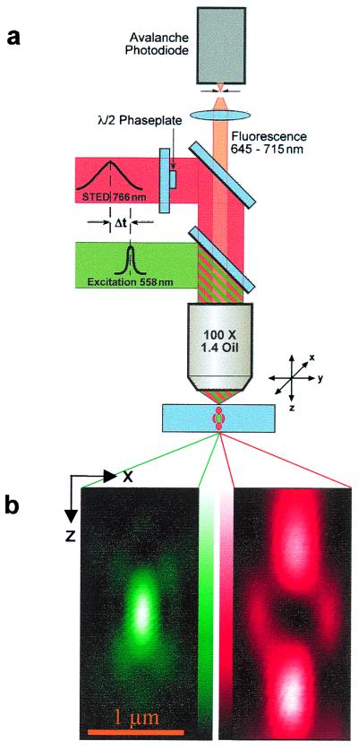
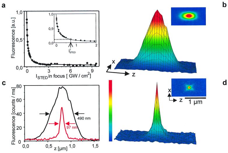
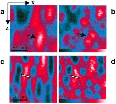
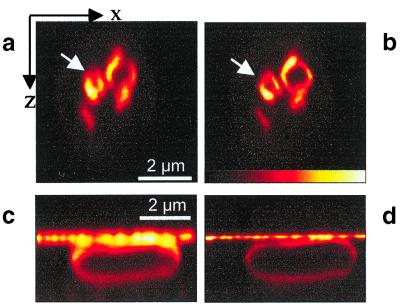
The diffraction barrier responsible for a finite focal spot size and limited resolution in far-field fluorescence microscopy has been fundamentally broken. This is accomplished by quenching excited organic molecules at the rim of the focal spot through stimulated emission. Along the optic axis, the spot size was reduced by up to 6 times beyond the diffraction barrier. The simultaneous 2-fold improvement in the radial direction rendered a nearly spherical fluorescence spot with a diameter of 90-110 nm. The spot volume of down to 0.67 attoliters is 18 times smaller than that of confocal microscopy, thus making our results also relevant to three-dimensional photochemistry and single molecule spectroscopy. Images of live cells reveal greater details.
Figures
Comment in
-
Shattering the diffraction limit of light: a revolution in fluorescence microscopy?Proc Natl Acad Sci U S A. 2000 Aug 1;97(16):8747-9. doi: 10.1073/pnas.97.16.8747. Proc Natl Acad Sci U S A. 2000. PMID: 10922028 Free PMC article. No abstract available.
-
The limits of light.Nat Rev Mol Cell Biol. 2010 Oct;11(10):678. doi: 10.1038/nrm2989. Nat Rev Mol Cell Biol. 2010. PMID: 20861874 No abstract available.
References
-
- Pawley J. Handbook of Biological Confocal Microscopy. New York: Plenum; 1995.
-
- Birks J B. Photophysics of Aromatic Molecules. London: Wiley Interscience; 1970.
-
- Hell S W, Wichmann J. Opt Lett. 1994;19:780–782. - PubMed
-
- Hell S W. In: Topics in Fluorescence Spectroscopy. Lakowicz J R, editor. Vol. 5. New York: Plenum; 1997. pp. 361–422.
Publication types
MeSH terms
Substances
LinkOut - more resources
Full Text Sources
Other Literature Sources
