. 2000 Jul 18;97(15):8403-8.
doi: 10.1073/pnas.97.15.8403.

Distinct spectra of somatic mutations accumulated with age in mouse heart and small intestine

Affiliations

Distinct spectra of somatic mutations accumulated with age in mouse heart and small intestine

M E Dollé et al. Proc Natl Acad Sci U S A. .

Abstract

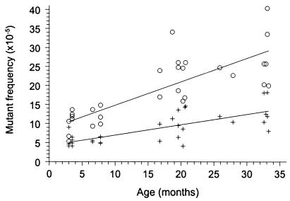
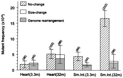
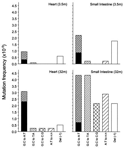
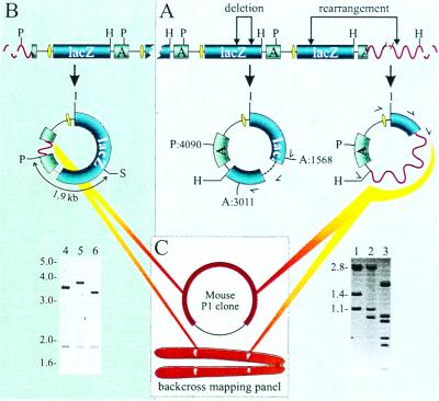
Somatic mutation accumulation has been implicated as a major cause of cancer and aging. By using a transgenic mouse model with a chromosomally integrated lacZ reporter gene, mutational spectra were characterized at young and old age in two organs greatly differing in proliferative activity, i.e., the heart and small intestine. At young age the spectra were nearly identical, mainly consisting of G. C to A.T transitions and 1-bp deletions. At old age, however, distinct patterns of mutations had developed. In small intestine, only point mutations were found to accumulate, including G.C to T.A, G.C to C.G, and A.T to C.G transversions and G.C to A.T transitions. In contrast, in heart about half of the accumulated mutations appeared to be large genome rearrangements, involving up to 34 centimorgans of chromosomal DNA. Virtually all other mutations accumulating in the heart appeared to be G.C to A.T transitions at CpG sites. These results suggest that distinct mechanisms lead to organ-specific genome deterioration and dysfunction at old age.


Figures

Figure 1
Figure 1
Spontaneous lacZ mutant frequencies in small intestine (○) and heart (+) of 3- to 33-month-old mice.
Figure 2
Figure 2
Mean frequencies of no-change mutants and size-change mutants in mouse heart and small intestine of 3.3- and 32-month-old animals (n = 7). Error bars indicate standard deviations of the means. Significant age-related changes for no-change mutants were found in both heart (P = 0.0049) and small intestine (P = 0.0022). Age-related changes for size-change mutants were significant for heart (P = 0.0073), but not for small intestine (P = 0.61; Wilcoxon Rank-Sum test). The shaded area indicates the fraction of size-change mutants with one break point in the mouse genome (genome rearrangements). The mutation load per cell, determined on the basis of a 3,000-bp lacZ target locus and a 6 × 10⁹-bp diploid genome, is given in parentheses above each bar.
Figure 3
Figure 3
Point mutational spectra of heart and small intestine at young and old age. Bars indicate the frequency of each type of point mutation as indicated. The black areas in the G:C to A:T bars indicate the fraction of such mutations that had occurred at CpG sites.
Figure 4
Figure 4
(A) Breakpoint identification of size-change mutants. Internal lacZ deletion events (lane 2, no. 305) or rearrangements across the mouse flanking sequence (lane 3, no. 205) were recognized by their PstI (P) and AvaI (A) restriction patterns deviating from the wild-type pattern (lane 1, the 5,346-bp plasmid results in 2.8-, 1.4-, and 1.1-kb restriction fragment lengths). The multiple fragments seen in lane 3 are caused by PstI and/or AvaI restriction sites in the recovered mouse sequence. H indicates the unique HindIII restriction site. The single-sided curved arrows indicate the direction and the location of the primers used to sequence the breakpoints. (B) Cloning of the integration sites of pUR288 concatemers in C57BL/6 transgenic mice. PstI digestion yields outer plasmid copies with a disrupted ampicillin resistance gene, containing the flanking genomic mouse sequence until the first PstI site (see Molecular Characterization of Size-Change Mutants for details). Lanes 4 and 5 show the plasmid digests with the upstream flanking sequence of integration site A (on chromosome 3) and B (on chromosome 4), respectively. Lane 6 shows the wild-type pattern (3.4- and 1.9-kb fragments). S indicates the unique SacI restriction site. (C) Strategy for determining the size of mouse sequence mutations. Mouse sequences recovered in mutant plasmids first were tested for their presence on P1 clones containing the integration sites. When these results were negative, a backcross panel was used to map the location of the recovered mouse fragment genetically (see Molecular Characterization of Size-Change Mutants for details).

References

    1. Yu C E, Oshima J, Fu Y H, Wijsman E M, Hisama F, Alisch R, Matthews S, Nakura J, Miki T, Ouais S, et al. Science. 1996;272:258–262. - PubMed
    1. Shen J C, Gray M D, Oshima J, Kamath-Loeb A S, Fry M, Loeb L A. J Biol Chem. 1998;273:34139–34144. - PubMed
    1. Yamagata K, Kato J, Shimamoto A, Goto M, Furuichi Y, Ikeda H. Proc Natl Acad Sci USA. 1998;95:8733–8738. - PMC - PubMed
    1. Fukuchi K, Martin G M, Monnat R J., Jr Proc Natl Acad Sci USA. 1989;86:5893–5897. - PMC - PubMed
    1. Turker M S, Martin G M. In: Principles of Geriatric Medicine and Gerontology. 4th Ed. Hazzard W R, Blass J P, Ettinger W H Jr, Halter J B, Ouslander J G, editors. New York: McGraw–Hill; 1999. pp. 21–44.

Publication types

LinkOut - more resources