. 2000 Jul 18;97(15):8542-7.
doi: 10.1073/pnas.97.15.8542.

Epidermal growth factor-induced nuclear factor kappa B activation: A major pathway of cell-cycle progression in estrogen-receptor negative breast cancer cells

Affiliations

Epidermal growth factor-induced nuclear factor kappa B activation: A major pathway of cell-cycle progression in estrogen-receptor negative breast cancer cells

D K Biswas et al. Proc Natl Acad Sci U S A. .

Abstract

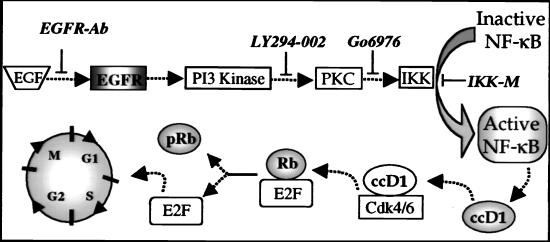
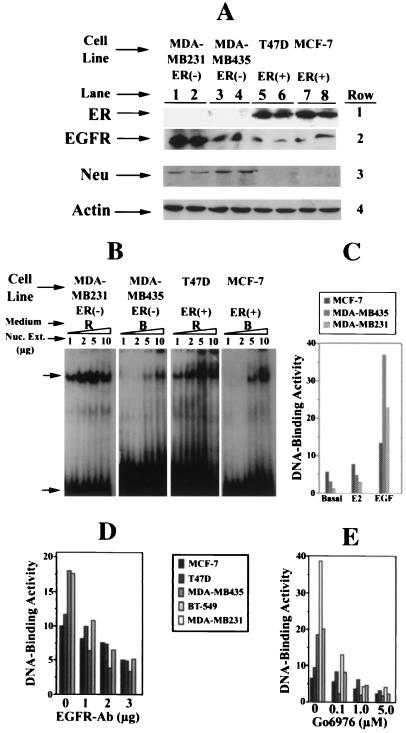
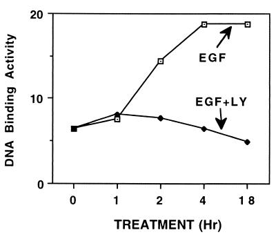
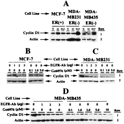
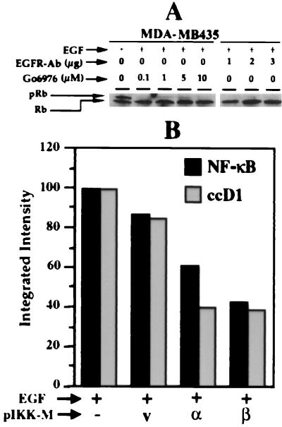
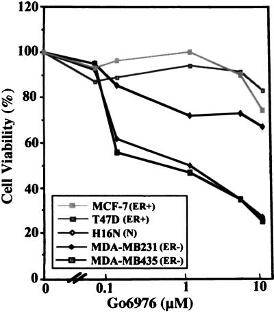
The epidermal growth factor (EGF) family of receptors (EGFR) is overproduced in estrogen receptor (ER) negative (-) breast cancer cells. An inverse correlation of the level of EGFR and ER is observed between ER- and ER positive (+) breast cancer cells. A comparative study with EGFR-overproducing ER- and low-level producing ER+ breast cancer cells suggests that EGF is a major growth-stimulating factor for ER- cells. An outline of the pathway for the EGF-induced enhanced proliferation of ER- human breast cancer cells is proposed. The transmission of mitogenic signal induced by EGF-EGFR interaction is mediated via activation of nuclear factor kappaB (NF-kappaB). The basal level of active NF-kappaB in ER- cells is elevated by EGF and inhibited by anti-EGFR antibody (EGFR-Ab), thus qualifying EGF as a NF-kappaB activation factor. NF-kappaB transactivates the cell-cycle regulatory protein, cyclin D1, which causes increased phosphorylation of retinoblastoma protein, more strongly in ER- cells. An inhibitor of phosphatidylinositol 3 kinase, Ly294-002, blocked this event, suggesting a role of the former in the activation of NF-kappaB by EGF. Go6976, a well-characterized NF-kappaB inhibitor, blocked EGF-induced NF-kappaB activation and up-regulation of cell-cycle regulatory proteins. This low molecular weight compound also caused apoptotic death, predominantly more in ER- cells. Thus Go6976 and similar NF-kappaB inhibitors are potentially novel low molecular weight therapeutic agents for treatment of ER- breast cancer patients.


Figures

Figure 1
Figure 1
A proposed pathway of EGF-induced cell proliferation of ER− breast cancer cells. The interaction of EGF with EGFR and the proposed downstream events of NF-κB activation and cell-cycle progression are schematically shown. The shaded molecules are monitored. The sites of action of agents used to block individual steps of this pathway, such as EGF–EGFR interaction by EGFR-Ab, PI3 kinase (phosphotidylinositol 3-kinase) activity by Ly294–002, PKC (protein kinase C) activity by G06976, and IKK activity by IKK-M (dominant-negative mutant of IKK) are shown. The role of G1-specific cell-cycle regulatory proteins, ccD1 and phosphorylated retinoblastoma (pRb), and unphosphorylated Rb is illustrated.
Figure 2
Figure 2
Levels of ER and EGFR family receptors and activation and inhibition of NF-κB in breast cancer cells. A shows the levels of ER, EGFR, Neu, and actin proteins in whole-cell extracts of ER− MDA-MB435 and MDA-MB231 and ER+ T47D and MCF-7 breast cancer cells in culture, as measured by Western blot analysis. Cells were grown in rich (R) medium to 90–95% confluency, whole-cell extracts were prepared (6), and 50 μg of protein in samples (in duplicate, designated by numerals under each cell line) was subjected to Western blot analysis and immunodetected with anti-ER-antibody Sc-543 (row 1) (42). The same blot was stripped and reused for detection of EGFR with anti-EGFR antibody Sc-03-G (row 2). Row 3 shows the level of Neu detected similarly with the anti-Neu antibody (Sc 284 G), and row 4 shows the levels of actin in the same samples as determined by reprobing the same blot with antiactin antibody, which serves as a loading control. These determinations were made three times, and results of one experiment are shown here. B shows the level of 32P-DNA-binding activity of NF-κB in the indicated amounts (protein) of nuclear extracts from ER− MDA-MB231 and ER+ MCF-7 cells grown in rich medium (R) or basal medium (B), as measured by EMSA (–39). The retarded specific NF-κB-32P-DNA complex is indicated by the upper arrow, and the free 32P-DNA (NF-κB–oligonucleotide) is indicated by the bottom arrow. C shows stimulation of NF-κB-32P-DNA-binding activity by E2 and EGF. The binding activity (numerals on the y axis) represents integrated intensity of the autoradiographic signals quantitated, as described in Materials and Methods. The ER− MDA-MB231 and MDA-MB435 and ER+ MCF-7 cells were plated in 25 ml of rich medium in 150-mm tissue culture dishes. Forty-eight hours later, the medium was removed, and cells were washed with basal medium (B) and replenished with 25 ml of the same medium. Seventy-two hours later, the medium was removed and replenished with 25 ml of basal medium, and cells were grown for an additional 12 h in the presence of either E2 (10−6 M) or EGF (12 ng/ml). Nuclear extracts from the treated and control cells were prepared (40), and NF-κB-32P-DNA-binding activities in 5 μg of nuclear extracts of these samples were measured by EMSA. One of four such experiments is reported here. D shows the NF-κB-32P-DNA-binding activity in nuclear extracts (5 μg) from the four breast cancer cell lines grown in basal medium plus EGF (12 ng/m) and indicated amounts of anti-EGFR-antibody per 10 ml of basal medium for 12 h. Growth and treatment conditions of the cells were the same as described in C. NF-κB-32P-DNA-binding activity was determined in nuclear extracts from two ER− MDA-MB-435 and MDA-MB231 and two ER+, MCF-7, T47D cells by EMSA and quantitated, as described above. E shows similar analysis for the determination of NF-κB-32P-DNA-binding activity in nuclear extracts of cells treated with indicated concentrations of G06976. Growth and treatment conditions of cells are the same as described in C. Nuclear extracts from three ER− and two ER+ cells were prepared and subjected to EMSA. Quantitation of the autoradiographic signals of the NF-κB-32P-DNA complex was the same as described above.
Figure 3
Figure 3
Kinetics of EGF-induced activation of NF-κB and inhibition by Ly294–002. The growth of ER− MDA-MB231, nuclear extract preparation, conditions for EGF treatment (12 ng/ml), and measurements of NF-κB-32P-DNA-binding activity are the same as described in Fig. 2C. The nuclear extracts from cells treated with EGF alone (indicated by EGF) for the specified time period or EGF and Ly294–002 (100 nM) together (indicated by EGF + LY) were prepared and similarly analyzed.
Figure 4
Figure 4
Level of ccD1 in EGF-treated cells and inhibition by EGFR-Ab and Go6976. A shows the levels of ccD1 in EGF (12 ng/ml) and E2 (10−6 M)-treated ER− and ER+ breast cancer cells. Growth conditions of the cells were the same as described in the legend to Fig. 2. The ER− MDA-MB231 and MDA-MB435 and ER+ MCF-7 cells were grown in basal medium in the presence of EGF (12 ng/ml) or E2 (10−6 M) for 12 h. Whole-cell extracts were prepared, and their level of ccD1 protein in 50 μg was determined by Western blot analysis. The membranes with blot-transferred fractionated proteins were then subjected to detection with anti-ccD1-antibody (Sc 8396) and enhanced chemiluminescence immunodetection system, as described (42). The same blot was stripped and reprobed with antiactin antibody, which serves as loading control. These experiments were repeated three times. The levels of ccD1 (row 1) in EGF-stimulated ER+ MCF-7 (B), ER− MDA-MB-231 (C), and ER− MDA-MB-435 (D) cells, simultaneously treated with indicated amounts of EGFR-Ab (micrograms per 10 ml) or Go6976 (μM) are shown. Fifty micrograms of whole cell extract proteins is used in each lane. Duplicate samples were analyzed in the case ER− MDA-MB-435 cells (D). Lanes designated by Actin (row 2) (A–D) represent the results of reprobing of each of these membranes with antiactin-Ab, which served as loading controls.
Figure 5
Figure 5
(A) Level of pRb protein in EGF, EGFR-Ab, and Go6976-treated ER− MDA-MB435 cells. Cells were grown in basal medium in the presence of EGF (12 ng/ml) and EGFR-Ab or Go6976 at the indicated concentrations for 12 h. The level of pRb and underphosphorylated Rb (Rb) in 50 μg of whole-cell extracts was detected by Western blot analysis and immunodetection with anti-Rb-antibody, as described above. (B) NF-κB DNA-binding activity and ccD1 level in IKK-M-transfected ER− MDA-MB435 cells. Results presented demonstrate the levels of NF-κB activation and ccD1 in EGF-treated ER− MDA-MB435 cells transiently transfected with dominant-negative IKK-α (α), IKK-β (β) mutants (IKK-M), and vector control (v) (53). Growth conditions and EGF treatment are as described in A. Cells were transfected with indicated plasmids (10 μg) by using Superfect Transfection Reagent (Qiagen, Chatsworth, CA) following the protocol of the supplier. Cells in fresh basal medium (B) were grown for an additional 48 h after transfection. EGF was added 4 h before harvesting, nuclear extracts were prepared, and the level of NF-κB-binding activity and ccD1 was measured as described above.
Figure 6
Figure 6
Sensitivity of ER− and ER+ breast cancer cell lines to Go6976. Sensitivity to Go6976 of ER− MDA-MB435 and MDA-MB231 and ER+ MCF-7 and T47D breast cancer cells and the immortalized normal human mammary epithelial cells H16N (54) grown in rich medium was determined by measurement of the number of viable cells by 3-(4,5-dimethylthiazol-2-yl)-2,5-diphenyl tetrazolium bromide assay after treatment with different concentrations of the drug for 7 days (43). Details of growth conditions are described in Materials and Methods. Results of the average of duplicate samples of one of the three experiments are shown here. The number of cells in drug-treated samples was expressed as percent of the untreated control, designated as 100%.

References

    1. Hortobagyi G N. N Engl J Med. 1998;339:974–984. - PubMed
    1. Brown M. Hematol Oncol Clin North Am. 1994;8:101–112. - PubMed
    1. Jensen E V. Breast Cancer Res Treat. 1981;26:2319–2326. - PubMed
    1. Jordan V C. Breast Cancer Res Treat. 1995;36:267–285. - PubMed
    1. Smith D F, Toft D O. Mol Endocrinol. 1993;7:4–11. - PubMed

Publication types