. 2000 Jul 18;97(15):8728-33.
doi: 10.1073/pnas.97.15.8728.

Dissociation of response conflict, attentional selection, and expectancy with functional magnetic resonance imaging

Affiliations

Dissociation of response conflict, attentional selection, and expectancy with functional magnetic resonance imaging

B J Casey et al. Proc Natl Acad Sci U S A. .

Abstract

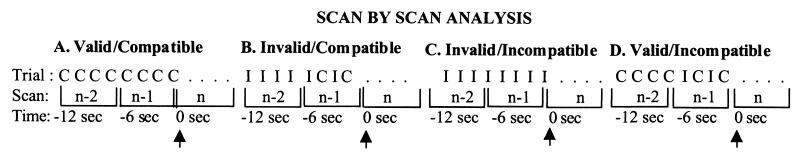
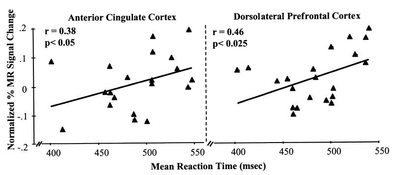
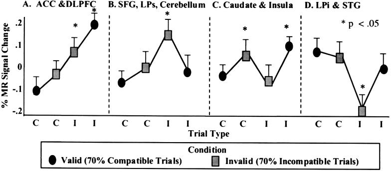
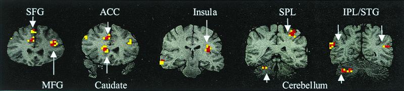
Two different attentional networks have been associated with visuospatial attention and conflict resolution. In most situations either one of the two networks is active or both are increased in activity together. By using functional magnetic resonance imaging and a flanker task, we show conditions in which one network (anterior attention system) is increased in activity whereas the other (visuospatial attention system) is reduced, showing that attentional conflict and selection are separate aspects of attention. Further, we distinguish between neural systems involved in different forms of conflict. Specifically, we dissociate patterns of activity in the basal ganglia and insula cortex during simple violations in expectancies (i.e., sudden changes in the frequency of an event) from patterns of activity in the anterior attention system specifically correlated with response conflict as evidenced by longer response latencies and more errors. These data provide a systems-level approach in understanding integrated attentional networks.


Figures

Figure 1
Figure 1
Post hoc scan-by-scan analyses were performed on brain regions identified as having significant MR signal change with the omnibus ANOVA for correct trials. Each 6-sec scan consisted of four behavioral trials, so the four trials were collapsed and averaged. In order for the MR signal to stabilize within an experimental condition (valid vs. invalid condition), scans containing the trial type of interest [compatible (C) or incompatible (I) trials] were preceded by at least one scan of all four compatible trials (valid condition) or all four incompatible trials (invalid condition). Thus the scans of interest consisted of four compatible trials preceded by four compatible trials (A); two compatible trials preceded by four incompatible trials (B); four incompatible trials preceded by four incompatible trials (C); and two incompatible trials preceded by four compatible trials (D). Scans that were analyzed (indicated by the arrows) consisted of those occurring 6 sec after the scan of interest to adjust for the typical 5- to 6-sec delay in peak of the hemodynamic response.
Figure 2
Figure 2
Percentage change in normalized MR signal intensity for the anterior cingulate cortex and dorsolateral prefrontal cortex and plotted as a function of mean reaction times for each subject on correct compatible and incompatible trials for the valid and invalid conditions.
Figure 3
Figure 3
Depiction of the four patterns of percentage change in normalized MR signal intensity as a function of compatible (C) and incompatible (I) trial type within the valid (70% compatible trials) and invalid (70% incompatible trials) conditions for correct trials across subjects. (A) The first pattern was observed in the anterior cingulate cortex (ACC) and dorsolateral prefrontal cortex (DLPFC). Activity in these regions increased as a function of increasing interference from the flanker stimuli and were evidenced by increasing response latencies. (B) A second pattern of results involved the right superior frontal gyrus (SFG), superior parietal lobule (LPs) and portions of the right cerebellum. These regions increased in activity during incompatible trials after consecutive incompatible trials (invalid condition). (C) A third pattern involving the basal ganglia and left insula showed increases in activity to compatible trials after consecutive incompatible trials or incompatible trials embedded in mostly compatible trials. (D) Finally, an inferior parietal (LPi) region and the superior temporal gyrus (STG) showed an inverse relation in activity to that observed in LPs and SFG. Activity in this region decreased during incompatible trials, particularly when embedded among incompatible trials (invalid condition).
Figure 4
Figure 4
Location of brain activity by gyrus and Brodmann's areas in regions demonstrating the four different patterns of percentage change in MR signal intensity depicted in Fig. 3 and illustrated here in the coronal plane.

References

    1. Corbetta M, Miezin F M, Shulman G L, Petersen S E. J Neurosci. 1993;13:1202–1226. - PMC - PubMed
    1. Nobre A C, Sebestyen G N, Gitelman D R, Mesulam M M, Frackowiak R S, Frith C D. Brain. 1997;120:515–533. - PubMed
    1. Bushnell M C, Goldberg M E, Robinson D L. J Neurophysiol. 1981;46:755–771. - PubMed
    1. Coull J T, Nobre A C. J Neurosci. 1998;18:7426–7435. - PMC - PubMed
    1. Pardo J V, Pardo P, Janer K W, Raichle M E. Proc Natl Acad Sci USA. 1990;87:256–259. - PMC - PubMed

Publication types

LinkOut - more resources