. 2000 Aug 1;97(16):9180-5.
doi: 10.1073/pnas.130196697.

Inhibition of growth and metastatic progression of pancreatic carcinoma in hamster after somatostatin receptor subtype 2 (sst2) gene expression and administration of cytotoxic somatostatin analog AN-238

Affiliations

Inhibition of growth and metastatic progression of pancreatic carcinoma in hamster after somatostatin receptor subtype 2 (sst2) gene expression and administration of cytotoxic somatostatin analog AN-238

N Benali et al. Proc Natl Acad Sci U S A. .

Abstract

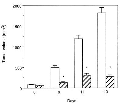
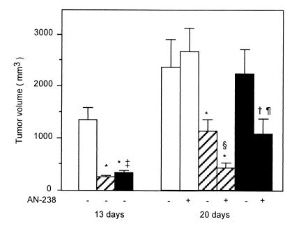
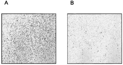
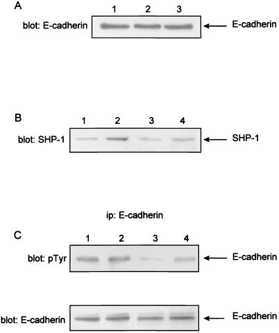
The sst2 somatostatin receptor mediates the antiproliferative effects of somatostatin analogs. The present study demonstrates that stable expression of sst2 in the hamster pancreatic cancer cells PC-1 and PC-1.0 activates an autocrine negative loop leading to an in vitro inhibition of cell proliferation. In vivo studies conducted in Syrian golden hamsters after orthotopic implantation of PC-1.0 cells showed that both tumor growth and metastatic progression of allografts containing 100% of sst2-expressing cells were significantly inhibited for up to 20 days after implantation, as compared with control allografts that did not express sst2. A local antitumor bystander effect was observed after induction of mixed tumors containing a 1:3 ratio of sst2-expressing cells to control cells. Tumor volume and incidence of metastases of mixed tumors were significantly reduced at day 13 post implantation. This effect decreased with time as at day 20, growth of mixed tumors was similar to that of control tumors. After administration of the cytotoxic somatostatin conjugate AN-238 on day 13, antitumor bystander effect observed in mixed tumors was significantly extended to day 20. We also observed that in vitro invasiveness of sst2-expressing PC-1.0 cells was significantly reduced. Tyrosine dephosphorylation of E-cadherin may participate in restoring the E-cadherin function, reducing in turn pancreatic cancer cell motility and invasiveness. This dephosphorylation depends on the tyrosine phosphatase src homology 2-containing tyrosine phosphatase 1 (SHP-1) positively coupled to sst2 receptor. The inhibitory effect of sst2 gene expression on pancreatic cancer growth and invasion combined with chemotherapy with targeted cytotoxic somatostatin analog administration provides a rationale for a therapeutic approach to gene therapy based on in vivo sst2 gene transfer.


Figures

Figure 1
Figure 1
Expression of human sst2 in hamster pancreatic cancer cells results in an autocrine negative loop. (A and B) PC1.0 cells stably expressing or not human sst2 receptor were cultured in 10-cm diameter dishes for 48 h in medium containing 5% FCS. Total RNA was extracted, and RT-PCR analysis was performed from clonal cell lines containing mock vector (1) or expressing human sst2 (2). The PCR products resulting from specific primers for human sst2 (A, 1107 pb), for preprosomatostatin (B, 487 pb), and β actin (A and B, 517 pb) were analyzed on polyacrylamide gels after ethidium bromide staining. M, DNA size marker (PGEM markers; Promega). RT-PCR carried out in the absence of reverse transcriptase during RT procedure were negative. Results are representative of two separate experiments. (C) Cells (4 × 10⁴ per 35-mm diameter dish) were grown in medium supplemented with 5% FCS. After 16-h attachment phase, cells were cultured in serum-free medium for 3 days. Cell growth was measured at the indicated times by cell counting. Results are expressed as the cell number per dish (mean ± SE) and are representative of two separate experiments in triplicate (□, PC-1.0 cells; ■, PC-1.0/sst2 cells; Δ, PC-1 cells; and ▴, PC-1/sst2 cells).
Figure 2
Figure 2
Effect of human sst2 expression on pancreatic cancer growth in hamsters. A total of 5 × 10⁵ PC-1.0 (open bars) or PC-1.0/sst2 (hatched bars) cells resuspended in 0.1 ml FCS-free RPMI medium 1640 were injected into the tail of the pancreas of Syrian golden hamsters (Exp. 1). The tumor's size was measured after laparatomy at indicated days post cell implantation. Results are means ± SE from four animals per group and per day of examination and are representative of two separate experiments (*, P < 0.001).
Figure 3
Figure 3
Effects of a single i.v. injection of cytotoxic somatostatin conjugate AN-238 on primary pancreatic cancer growth in hamsters. PC-1.0 (open bars), PC-1.0/sst2 (hatched bars), and a mixture of PC-1.0/sst2 and PC-1.0 cells (ratio of PC-1.0/sst2 to PC-1.0 = 25:75; filled bars) were inoculated orthotopically into the tail of the pancreas of hamster (5 × 10⁵ per site). Tumor volumes were measured at day 13 as described for Exp. 2 and at day 20 in group that did (+) or did not (−) receive single i.v. injections of 100 nmol/kg of AN-238 at day 13 according to Exp. 3. Values are from 20 animals at day 13 from four separate experiments and from 10 animals per group at day 20 from two separate experiments. Difference from control tumors (open bar): *, P < 0.001; †, P < 0.01. Difference between sst2 and mixed tumor volume at 13 days: ‡, P < 0.05. Difference with tumor volume at 20 days in untreated animals: §, P < 0.01; ¶, P < 0.05.
Figure 4
Figure 4
Effect of sst2 expression on in vitro invasivenes of pancreatic cancer cells. Cells were plated in chamber containing a Matrigel-coated filter at its base. The chambers were immersed in RPMI medium 1640 with 1% of BSA for 30 min at 37°C. After 48-h incubation, the noninvasive cells, which remain on the topside of the filter, were removed, and the invasive cells, which attach themselves to the underside of the filter, were stained with 0.2% crystal violet after fixing with 3% paraformaldehyde. The figure represents a photograph at a low magnification (×40) of filters with stained cells (A, PC-1.0 cells; B, PC-1.0/sst2 cells) and is representative of four separate experiments.
Figure 5
Figure 5
Effect of sst2 expression on E-cadherin protein expression and tyrosine dephosphorylation in pancreatic cancer cells. (A) Cells were cultured in 10-cm diameter dishes in medium containing 5% FCS. After a 12-h attachment phase, cells were cultured in serum-free medium for 24 h. Soluble proteins were subjected to SDS/polyacrylamide gel electrophoresis and immunoblotted (blot E-cadherin) with anti-E-cadherin Abs. Lane 1, PC1.0/neo cells; lane 2, PC-1.0 cells; and lane 3, PC1.0/sst2 cells. The arrow indicates the position of E-cadherin (120 kDa). Results are representative of three independent experiments. (B and C) PC-1.0 and PC-1.0/sst2 cells transiently transfected or not with the pcDNA3/C453S-SHP-1 vector (dominant negative SHP-1 mutant) were cultured in 100-mm dishes (7 × 10⁵) cells/dish in RPMI medium 1640 supplemented with 5% FCS for 18 h. Cells were thereafter solubilized and immunoprecipitated or not with E-cadherin Abs. Solubilized proteins were resolved through 7.5% SDS/polyacrylamide gels and immunoblotted with anti-SHP-1 Abs (B, blot SHP-1). Immunoprecipitated proteins (ip: E-cadherin) were immunoblotted with anti-phosphotyrosine (C, blot: p-Tyr) or anti-E-cadherin (C, blot: E-cadherin, reblotting experiment). The arrow indicates the position of E-cadherin phosphorylation (120 kDa). Lanes 1, PC-1.0 cells; lanes 2, PC-1.0-SHP-1/C453S cells; lanes 3, PC-1.0/sst2 cells; lanes 4, PC-1.0/sst2-SHP-1/C453S cells. The figure is representative of three independent experiments.

References

    1. Lamberts S W, Krenning E P, Reubi J C. Endocr Rev. 1991;12:450–482. - PubMed
    1. Pollak M N, Schally A V. Proc Soc Exp Biol Med. 1998;217:143–152. - PubMed
    1. Arnold R, Frank M. Digestion. 1996;57, Suppl. 1:69–71. - PubMed
    1. Hoyer D, Bell G I, Berelowitz M, Epelbaum J, Feniuk W, Humphrey P P, AM, O C, Patel Y C, Schonbrunn A, Taylor J E, Reisine T. Trends Pharmacol Sci. 1995;16:86–88. - PubMed
    1. Buscail L, Esteve J P, Saint-Laurent N, Bertrand V, Reisine T, AM, O C, Bell G I, Schally A V, Vaysse N, Susini C. Proc Natl Acad Sci USA. 1995;92:1580–1584. - PMC - PubMed

Publication types

MeSH terms