. 2000 Jul;106(2):299-308.
doi: 10.1172/JCI9860.

Loss of function and inhibitory effects of human CSX/NKX2.5 homeoprotein mutations associated with congenital heart disease

Affiliations

Loss of function and inhibitory effects of human CSX/NKX2.5 homeoprotein mutations associated with congenital heart disease

H Kasahara et al. J Clin Invest. 2000 Jul.

Abstract

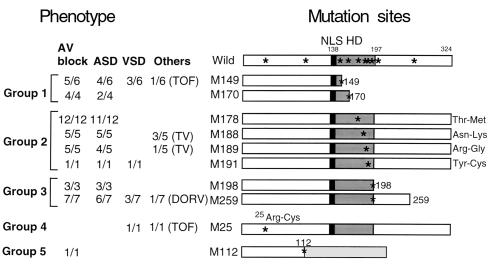
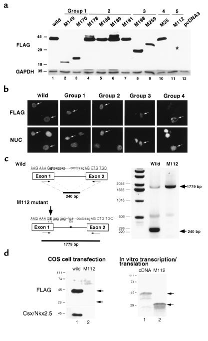
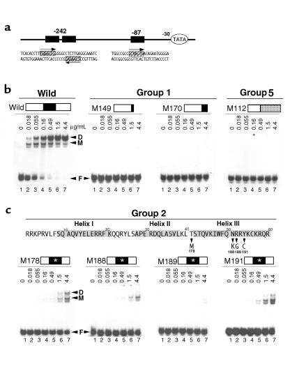
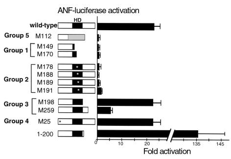
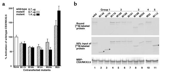
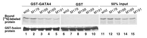
CSX/NKX2.5 is an evolutionarily conserved homeodomain-containing (HD-containing) transcription factor that is essential for early cardiac development. Recently, ten different heterozygous CSX/NKX2.5 mutations were found in patients with congenital heart defects that are transmitted in an autosomal dominant fashion. To determine the consequence of these mutations, we analyzed nuclear localization, DNA binding, transcriptional activation, and dimerization of mutant CSX/NKX2.5 proteins. All mutant proteins were translated and located to the nucleus, except one splice-donor site mutant whose protein did not accumulate in the cell. All mutants that had truncation or missense mutations in the HD had severely reduced DNA binding activity and little or no transcriptional activation function. In contrast, mutants with intact HDs exhibit normal DNA binding to the monomeric binding site but had three- to ninefold reduction in DNA binding to the dimeric binding sites. HD missense mutations that preserved homodimerization ability inhibited the activation of atrial natriuretic factor by wild-type CSX/NKX2.5. Although our studies do not characterize the genotype-phenotype relationship of the ten human mutations, they identify specific abnormalities of CSX/NKX2.5 function essential for transactivation of target genes.


Figures

Figure 1
Figure 1
Diagram of CSX/NKX2.5 cDNA with the location of ten mutation sites identified in congenital heart disease. Ten mutation sites (asterisks in Wild) were divided into five groups based on the predicted protein structure: nonsense mutation in the HD (group 1: M149 and M170); missense mutation in the HD (group 2: M178, M188, M189, and M191); premature termination after HD (group 3: M198 and M259); 25Arg-Cys missense mutation (group 4: M25); and mutation at the intron-splicing donor site (group 5: M112). Phenotypes observed in patients are listed on the left. For example, “11/12” indicates that 11 patients show the phenotype among 12 patients examined. These mutation sites were mapped on CSX/NKX2.5 cDNA, which encodes 324 amino acids including 60 amino acids of HD (shaded box). Nuclear localization signal at the NH2-terminus of the HD is indicated (black box). Predicted translated product of M112 mutation in splicing donor site is indicated with a light gray box. AV block, atrioventricular conduction block; ASD, atrial septal defect; VSD, ventricular septal defect; TOF, tetralogy of Fallot; TV, tricuspid valve abnormality; DORV, double outlet right ventricle; NLS, nuclear localization signal; HD, homeodomain.
Figure 2
Figure 2
Expression of translated products in cells: intron-splicing defect in M112 mutant resulted in poor protein accumulation. (a) Wild-type and mutant CSX/NKX2.5 expression vectors were transfected into COS 7 cells, and the protein expression was examined by Western blotting using anti-FLAG Ab approximately 24 hours after transfection (FLAG, top). All mutant proteins except M112 (lane 11, asterisk indicates the expected molecular weight of M112 protein) were detected at the expected molecular weight. GAPDH expression in each lane was also shown (GAPDH, bottom). (b) Wild-type and mutant CSX/NKX2.5 proteins accumulated in the nucleus colocalizing with Hoechst nuclear staining (NUC, lower panels). The results presented are wild-type, group 1 (M170), group 2 (M191), group 3 (M198), and group 4 (M25). (c) G→T substitution (large arrow) identified in M112 mutant on the CSX/NKX2.5 splicing donor site was examined by RT-PCR. RNA-purified form COS 7 cells transfected with the wild-type and M112 mutant of CSX/NKX2.5 were amplified with two primers spanning the intron. In the wild-type transfectant, the intron was spliced out, resulting in the generation of 240-bp product, whereas in the mutant transfectant, G→T substitution of the first codon of the intron splicing site abolished the normal splicing, resulting in generation of 1,779-bp product. CSX/NKX2.5 protein was encoded by the two exons represented. (d) Translated products were examined approximately 24 hours after transfection by Western blotting using anti-FLAG mAb (top) and anti-Csx/Nkx2.5 mAb (bottom). Wild-type CSX/NKX2.5 gene was translated into approximately 42-kDa protein and was detected with anti-FLAG and anti-Csx/Nkx2.5 mAb (lane 1). In contrast, M112 mutant protein that is expected to migrate approximately 29 kDa was not detected in the cell lysate (lane 2). In in vitro transcription and translation system, cDNA produced approximately 42-kDa protein (lane 1), and M112 genomic construct produced about 29-kDa protein (lane 2).
Figure 3
Figure 3
DNA binding affinity of group 1, 2, and 5 mutant proteins versus wild-type CSX/NKX2.5. (a) Sequence of two consensus CSX/NKX2.5 binding sites (–242 bp and –87 bp sites) in rat ANF promoter; the paired binding sites in –242 bp site, and a single binding site in –87 bp site. (b) –242 bp site was used for the DNA binding assay (lane 1) mixed with threefold serially increased CSX/NKX2.5 fusion proteins (0.018–4.4 μg/mL of MBP-CSX/NKX2.5 fusion protein) (lanes 2–7). Wild-type CSX/NKX2.5 bound as a monomer (M) as well as a dimer (D) depending on the protein concentration. No shifted bands were observed in groups 1 and 5 (M149, M170, and M112). (c) The EMSA of the group 2 mutant proteins that have a single missense mutation in the HD. 178Thr-Met (M178) mutation was located just before the third helix, and 188Asn-Lys (M188), 189Arg-Gly (M189), and 191Tyr-Cys (M191) were located in the third helix. Two conserved amino acid mutations were identified: 188Asn (51Asn in HD) is conserved in all the HD protein that is directly bound to the major groove of DNA, and 191Tyr (54Tyr in HD) is conserved in all NK2 class homeoprotein. All four mutant proteins show dramatically reduced DNA binding affinity compared with the wild-type CSX/NKX2.5. D, dimer; M, monomer; F, free probe. Mutation sites of group 2 are indicated with white asterisks.
Figure 4
Figure 4
DNA binding affinity of the group 3 and 4 mutant proteins versus wild-type CSX/NKX2.5. Sequence of the native –242 bp site (top) and a mutated –242 bp binding site (bottom) in the ANF promoter. EMSA of group 3 (M198 and M259) and group 4 (M25, the mutation site is marked with an asterisk) protein compared with that of the wild-type CSX/NKX2.5. Proteins were mixed with probes containing either tandemly repeated binding sites (ad) or single binding site (eh). Lanes showing similar monomer/dimer ratios are indicated with asterisks in the top panels (ad). In all three mutant proteins, binding affinity as a dimer is reduced approximately 3- to 32 fold (bd versus a), whereas they show similar DNA binding affinity as wild-type to the single binding site (fh versus e). D, dimer; M, monomer; F, free probe.
Figure 5
Figure 5
Effect of ten mutations on transcriptional activation. 10T1/2 cells were transfected with pcDNA3 expression vectors encoding wild-type or each of ten mutations with the reporter gene ANF-luciferase. When the wild-type CSX/NKX2.5 was transfected with the ANF reporter gene, luciferase activity was increased 23-fold compared with cells transfected with the empty expression vector pcDNA3. M112, group 1, and group 2 expression vectors did not activate the ANF promoter. M25 and M198 transactivated the reporter gene similarly to the wild-type CSX/NKX2.5; however, M259 transactivated only 5.2-fold. Another COOH-terminus deletion mutant, CSX/NKX2.5(1–200), transactivated the reporter construct approximately 136-fold. Bars represent means ± SEM of at least three separate transfection assays done in duplicate.
Figure 6
Figure 6
Inhibition of ANF-luciferase transcriptional activity of wild-type CSX/NKX2.5 by group 1, 2, and 3 mutants but not by group 4 mutant. (a) Inhibition of transactivation of the ANF-luciferase reporter gene in the presence of both 1:1 and 2:1 ratio of M170, M189, M191, and M259 expression plasmid to wild-type expression plasmid. 10T1/2 cells were transiently transacted with 1.2 μg of ANF-luciferase(-638), 0.3 μg of RSV–β-galactosidase, 0.7 μg of wild-type CSX/NKX2.5 expression plasmid, and 0.7 μg (hatched bars) or 1.4 μg (filled bars) of mutant expression plasmid or empty pcDNA3 plasmid to adjust the total amount of plasmid. A moderate reduction of luciferase activity was observed in M170 mutants, and a further decrease of luciferase activity was detected in M189, M191, and M250 mutants. In contrast, M25 mutant increased luciferase activity. Results are presented as a percent of the ANF reporter activity when cotransfected with wild-type and mutant plasmid compared with that of the wild type alone (open bar). Bars represent means ± SEM of the means of at least three separate transfection assays done in duplicate. (b) Protein-protein interaction of wild-type CSX/NKX2.5 with mutant proteins. MBP-fused wild-type CSX/NKX2.5 protein was mixed with [35S]-labeled wild-type (lane 1) or ten mutants (lanes 2–11). After washing five times with binding buffer, the protein complexes were resolved on SDS-PAGE and autoradiographed (top panel). Group 1 (lanes 2 and 3) and group 5 (lane 11) mutants did not associate with MBP-CSX/NKX2.5, whereas group 2, 3, and 4 mutants associated with MBP-CSX/NKX2.5 (lanes 4–10). Fifty percent input of [35S]-labeled proteins are shown in the middle panel, and Coomassie blue–stained MBP-fused wild-type CSX/NKX2.5 proteins are shown in the bottom panel.
Figure 7
Figure 7
Interaction of group 2 mutants with GATA4 protein. [35S]-labeled wild-type CSX/NKX2.5 and four group 2 mutant proteins were mixed with GST-GATA4 protein (lanes 1–5) or GST alone (lanes 6–10). Bound labeled proteins were resolved on SDS-PAGE and autoradiographed (top panel). Fifty percent input of [35S]-labeled proteins is also shown. Coomassie blue–stained GST-GATA4 (lanes 1–5) or GST (lanes 6–10) fusion proteins are shown (bottom panel).

References

    1. Gehring WJ, Affolter M, Burglin T. Homeodomain proteins. Annu Rev Biochem. 1994;63:487–526. - PubMed
    1. Duboule, D. 1994. Guidebook to the homeobox genes. Oxford University Press. New York, New York, USA. 1–284.
    1. Damante G, et al. Sequence-specific DNA recognition by the thyroid transcription factor-1 homeodomain. Nucleic Acids Res. 1994;22:3075–3083. - PMC - PubMed
    1. Harvey RP. NK-2 homeobox genes and heart development. Dev Biol. 1996;178:203–216. - PubMed
    1. Gruschus JM, Tsao DH, Wang LH, Nirenberg M, Ferretti JA. Interactions of the vnd/NK-2 homeodomain with DNA by nuclear magnetic resonance spectroscopy: basis of binding specificity. Biochemistry. 1997;36:5372–5380. - PubMed

Publication types