

The first human case of Trichinella spiralis infection in Korea
- PMID: 10905075
- PMCID: PMC2721112
- DOI: 10.3347/kjp.2000.38.2.111
The first human case of Trichinella spiralis infection in Korea
Abstract
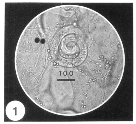
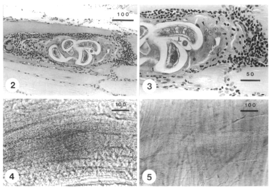
Three cases of human infection by Trichinella spiralis were first confirmed by detecting encysted larvae in the biopsied muscle in December 1997, in Korea. The patients were one 35- and two 39-year-old males residing in Kochang-gun, Kyongsangnam-do. They had a common past history of eating raw liver, spleen, blood and muscle of a badger, Meles meles melanogenys, and complained of high fever, facial and periorbital edema, and myalgia. Hematologic and biochemical examinations revealed leukocytosis and eosinophilia, and highly elevated levels of GOT, GPT, LDH and CPK. In the gastrocnemius muscle of a patient, roundly coiled nematode larvae were detected. The larvae measured 0.775-1.050 (av. 0.908) mm in length, and 0.026-0.042 (av. 0.035) mm in maximum width. The specific IgG antibody levels in three patients' sera were significantly higher when compared with those of normal controls. The patients were treated with flubendazole and albendazole for 15-30 days, and discharged at 13-34 days post-admission. From the above findings, it was confirmed that T. spiralis is present in Korea, and the badger plays a role of as the natural host.
Figures
References
-
- Despommier DD. Trichinella and Toxocara. In: Cox FEG, Kreier JP, Wakelin D, editors. Parasitology (Vol 5) of Topley & Wilson's Microbiology and Microbial Infections. 9th ed. London, UK: Arnold; 1998. pp. 597–602.
-
- Dworkin MS, Gamble HR, Zarlenga DS, Tennican PO. Outbreak of trichinellosis associated with eating cougar jerky. J Infect Dis. 1996;174:663–666. - PubMed
-
- Gould SE. In: Clinical pathology: diagnostic laboratory procedures, Trichinosis in man and animals. Gould SE, Thomas CC, editors. Illinois, USA: Springfield; 1970. pp. 191–221.
-
- Jongwutiwes S, Chantachum N, Kraivichian P, et al. First outbreak of human trichinellosis caused by Trichinella pseudospiralis. Clin Infect Dis. 1998;26:111–115. - PubMed
Publication types
MeSH terms
LinkOut - more resources
Full Text Sources
Miscellaneous
