. 2000 Aug;74(16):7230-7.
doi: 10.1128/jvi.74.16.7230-7237.2000.

The human cytomegalovirus 86-kilodalton major immediate-early protein interacts physically and functionally with histone acetyltransferase P/CAF

Affiliations

The human cytomegalovirus 86-kilodalton major immediate-early protein interacts physically and functionally with histone acetyltransferase P/CAF

L A Bryant et al. J Virol. 2000 Aug.

Abstract

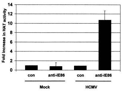
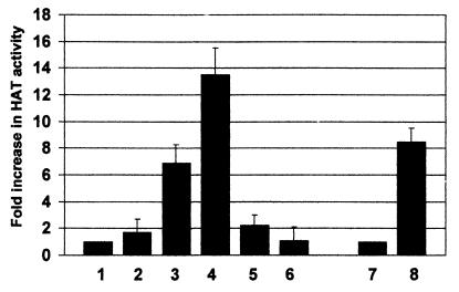
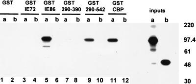
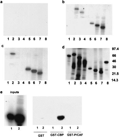
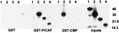
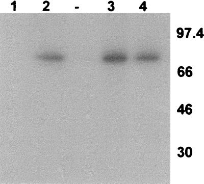
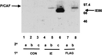
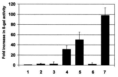
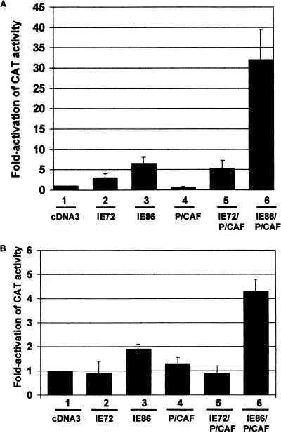
The major immediate-early proteins of human cytomegalovirus (HCMV) play a pivotal role in controlling viral and cellular gene expression during productive infection. As well as negatively autoregulating its own promoter, the HCMV 86-kDa major immediate early protein (IE86) activates viral early gene expression and is known to be a promiscuous transcriptional regulator of cellular genes. IE86 appears to act as a multimodal transcription factor. It is able to bind directly to target promoters to activate transcription but is also able to bridge between upstream binding factors such as CREB/ATF and the basal transcription complex as well as interacting directly with general transcription factors such as TATA-binding protein and TFIIB. We now show that IE86 is also able to interact directly with histone acetyltransferases during infection. At least one of these factors is the histone acetyltransferase CBP-associated factor (P/CAF). Furthermore, we show that this interaction results in synergistic transactivation by IE86 of IE86-responsive promoters. Recruitment of such chromatin-remodeling factors to target promoters by IE86 may help explain the ability of this viral protein to act as a promiscuous transactivator of cellular genes.


Figures

FIG. 1
FIG. 1
HAT activity coimmunoprecipitates with IE86 in infected cells. U373 cells were either mock infected (Mock) or infected with HCMV (HCMV) at approximately 5 PFU/cell; 24 h postinfection, cells were lysed and extracts were immunoprecipitated with a control monoclonal antibody to HCMV gB (con) or a monoclonal antibody to IE86 (anti-IE86). Immunocomplexes were then assayed for HAT activity. Data represent average fold increases in HAT activity over the corresponding control antibody immunoprecipitations from three independent experiments.
FIG. 2
FIG. 2
HAT activity coimmunoprecipitates with IE86 in transfected cells. U373 cells were transfected with pcDNA3 (bar 1), pcDNA3IE72 (bar 2), pcDNA3IE86 (bar 3), pcDNA3IE86 plus pCX-Flag-P/CAF (P/CAF) (bar 4), pcDNA3IE72P/CAF (bar 5), or P/CAF alone (bar 8). At 48 h posttransfection, cells were lysed and extracts were immunoprecipitated with E13 antibody, specific for HCMV IE72/IE86 (bars 1 to 5). An equivalent amount of extract from pcDNA3IE86+P/CAF-transfected cells was also immunoprecipitated with a monoclonal antibody to an HCMV late structural protein (Chemicon) as a control (bar 6). Data represent average fold increases in HAT activity over the HAT activity detected for pcDNA3 transfections from three independent experiments. Extracts from P/CAF-transfected cells were also immunoprecipitated with the control late HCMV antibody (bar 7) or an anti-Flag antibody, which detects the Flag epitope on P/CAF (lane 8), to act as a positive control for HAT detection. Immunocomplexes were then assayed for HAT activity. Data represent average fold increases in HAT activity over the HAT activity detected in the corresponding control antibody immunoprecipitations from three independent experiments.
FIG. 3
FIG. 3
P/CAF interacts with IE86 in vitro. In vitro-transcribed and -translated P/CAF (lanes a) or gelsolin (lanes b) was analyzed by GST fusion pull-down assays for the ability to bind GST, GST-IE72, GST-IE86, or GST-CBP beads. GST fusions to the 290–390 (GST 290–390) or 290–542 (GST 290–542) amino acid domain of IE86 were also analyzed. Inputs were 25% of the amount of IE86 or gelsolin used in each assay. Here and in subsequent figures, positions of marker proteins are indicated in kilodaltons.
FIG. 4
FIG. 4
Minimal domains of IE86 interact with P/CAF in vitro. GST (a), GST-CBP (b), and GST-P/CAF (c) beads were used as targets for GST pull-down assays using [35S]methionine-labeled IE72 (lanes 1), full-length IE86 (lanes 2), amino acids 1 to 390 (lanes 3), 1 to 290 (lanes 4), 290 to 579 (lanes 5), 290 to 542 (lanes 6), and 290 to 504 (lanes 7) of IE86, as well as gelsolin (lane 8). Input protein (25% of the amount of protein used in each assay) is shown in panel d. (e) GST, GST-CBP, and GST-P/CAF beads were used as targets in GST pull-down assays using [35S]methionine-labeled amino acids 1 to 85 (1) or 290 to 390 (2) of IE86; 25% of the amount of protein used in each assay is shown as inputs.
FIG. 5
FIG. 5
P/CAF and CBP require different domains of IE86 for interaction. GST, GST-P/CAF, and GST-CBP beads were used as targets for GST pull-down assays using [35S]methionine-labeled amino acids 290 to 579 (lane 2), 388 to 579 (lane 3), or 428 to 579 (lane 4) of IE86 and gelsolin (lane 1). Inputs represent 25% of the amount of protein used in each assay.
FIG. 6
FIG. 6
Interaction between P/CAF and IE86 requires no other eukaryotic protein. Bacterially expressed IE86 protein was labeled in vitro with 32P and used in GST pull-down assays to analyze binding to GST (lane 1), GST-P/CAF (lane 2), GST-TBP (lane 3), and GST-CBP (lane 4). A lane between lanes 2 and 3 was left unloaded to ensure no accidental overspill of the GST-TBP positive control sample.
FIG. 7
FIG. 7
IE86 and P/CAF interact in vivo. U2-OS cells were cotransfected with pcDNA3IE86 and pCX-Flag-P/CAF and labeled with [35S]methionine; 48 h posttransfection, cells were lysed in EBC buffer and extracts were subjected to double immunoprecipitation assays using a control anti-murine cyclin D1 monoclonal antibody (CON), an anti-IE antibody (IE), and an anti-Flag monoclonal antibody (FLAG) for primary immunoprecipitations (10). Complexes were washed and reimmunoprecipitated using the control (lanes a), anti-Flag (lanes b), and anti-IE (lanes c) antibodies for secondary (20) immunoprecipitations. Complexes were separated by SDS-PAGE and autoradiographed.
FIG. 8
FIG. 8
IE86 and P/CAF interact in yeast two-hybrid assays. The parental plasmid for expression in yeast of the DNA binding domain of GAL4 (pGBT10) was fused to P/CAF (pGBT:P/CAF) or IE86 (pGBT:IE86). The parental plasmid for expression in yeast of the GAL4 activation domain (pGAD425) was fused to IE86 (pGAD:IE86) or IE72 (pGAD:IE72). pGBT10 plus pGAD425 (bar 1), pGBT10 plus pGAD:IE86 (bar 2), pGBT:P/CAF plus pGAD425 (bar 3), pGBT:P/CAF plus pGAD:IE86 (bar 4), pGBT:IE86 plus pGAD:IE86 (bar 5), pGBT:P/CAF plus pGAD:IE72 (bar 6), and pVA3-1 plus pTD1-1 containing a GAL4 DNA binding domain murine p53 fusion protein and a GAL4 activation domain simian virus 40 T-antigen fusion, respectively (bar 7), were transformed into yeast, and β-galactosidase expression was measured in liquid culture. Data represent average fold increases in β-galactosidase activity over activity obtained from cotransformation with pGBT10 plus pGAD425 from three independent experiments.
FIG. 9
FIG. 9
IE86 and P/CAF synergistically activate transiently or stably transfected target promoters in transfection assays. (A) U373 cells were transiently transfected with TGFβ77MCAT together with pcDNA3 (bar 1), pcDNA3IE72 (bar 2), pcDNA3IE86 (bar 3), pCX-Flag-P/CAF (bar 4), pcDNA3IE72 and pCX-Flag-P/CAF (bar 5), or pcDNA3IE86 and pCX-Flag-P/CAF (bar 6). Data represent the average of three independent experiments. (B) U373 cells that had been stably transfected with TGFβ77CAT were transiently transfected with pcDNA3 (bar 1), pcDNA3IE72 (bar 2), pcDNA3IE86 (bar 3), pCX-Flag-P/CAF (bar 4), pcDNA3IE72 and pCX-Flag-P/CAF (bar 5), or pcDNA3IE86 and pCX-Flag-P/CAF (bar 6). Data represent the average of three independent experiments.

References

    1. Arlt H, Lang D, Gebert S, Stamminger T. Identification of binding sites for the 86-kilodalton IE2 protein of human cytomegalovirus within an IE2-responsive viral early promoter. J Virol. 1994;68:4117–4125. - PMC - PubMed
    1. Bannister A J, Kouzarides T. The CBP co-activator is a histone acetyltransferase. Nature. 1996;384:641–643. - PubMed
    1. Bannister A J, Kouzarides T. CBP-induced stimulation of c-Fos activity is abrogated by E1A. EMBO J. 1995;14:4758–4762. - PMC - PubMed
    1. Bannister A J, Oehler T, Wilhelm D, Angel P, Kouzarides T. Stimulation of c-Jun activity by CBP: c-Jun residues Ser63/73 are required for CBP induced stimulation in vivo and CBP binding in vitro. Oncogene. 1995;11:2509–2514. - PubMed
    1. Bresnahan W A, Albrecht T, Thompson E A. The cyclin E promoter is activated by human cytomegalovirus 86-kDa immediate early protein. J Biol Chem. 1998;273:22075–22082. - PubMed

Publication types

MeSH terms

Substances