. 2000 Aug;74(16):7338-48.
doi: 10.1128/jvi.74.16.7338-7348.2000.

Viral activation of interleukin-15 (IL-15): characterization of a virus-inducible element in the IL-15 promoter region

Affiliations

Viral activation of interleukin-15 (IL-15): characterization of a virus-inducible element in the IL-15 promoter region

N Azimi et al. J Virol. 2000 Aug.

Erratum in

  • J Virol 2002 Jul;76(13):6864

Abstract

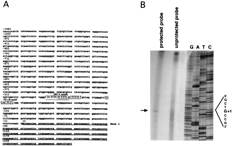
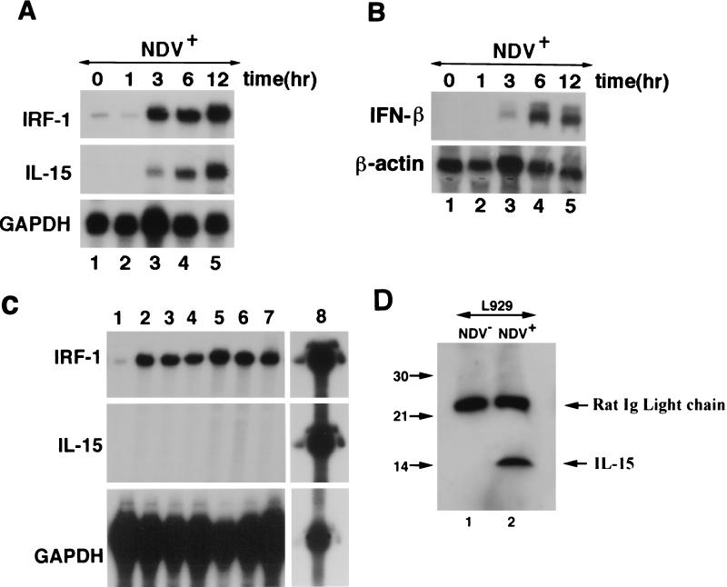
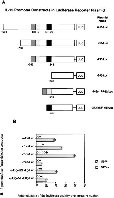
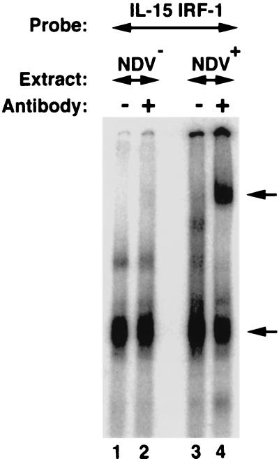
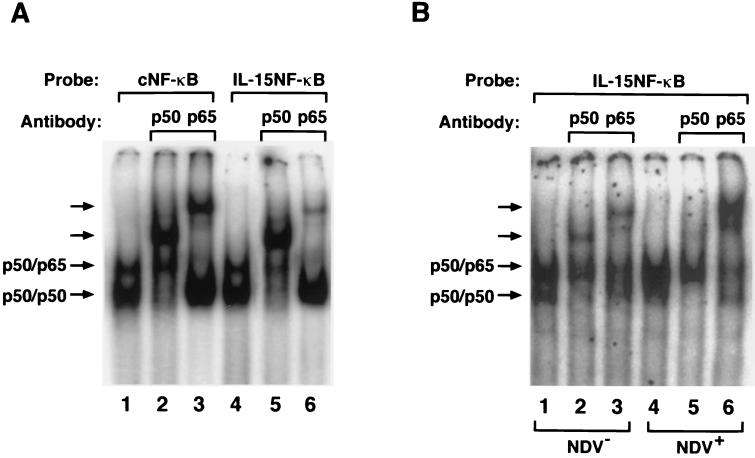
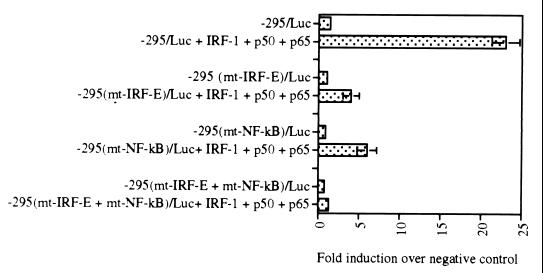
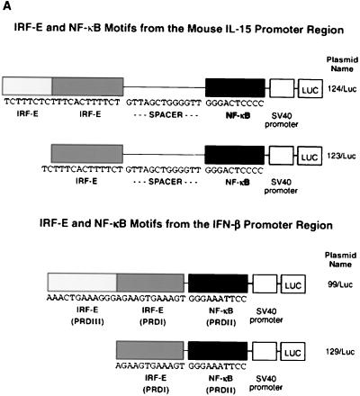
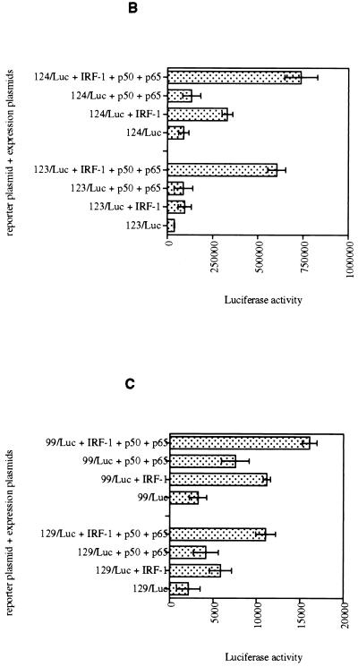
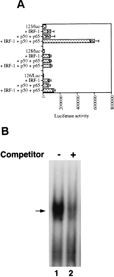
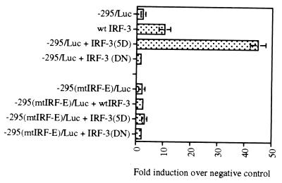
We identified an interferon regulatory factor motif (IRF-E) upstream of an NF-kappaB binding site in the interleukin-15 (IL-15) promoter. Since these two motifs are part of the virus-inducible enhancer region of the beta interferon promoter, we speculated that there might be similar responses of these two genes to stimuli such as viruses. To test this hypothesis, L929 cells were infected with Newcastle disease virus (NDV), which led to the induction of IL-15 mRNA and protein expression. Using IL-15 promoter-reporter deletion constructs, a virus-inducible region, encompassing IRF-E, NF-kappaB, and a 13-nucleotide sequence flanked by these two motifs, was mapped to the -295-to--243 position relative to the transcription initiation site. Using cotransfection studies, it was demonstrated that all three motifs were essential to achieve the maximum promoter activity induced by IRF-1 and NF-kappaB expression plasmids. The presence of a virus-inducible region in the IL-15 promoter suggests a role for IL-15 as a component of host antiviral defense mechanisms.


Figures

FIG. 1
FIG. 1
(A) Nucleotide sequence of the mouse IL-15 5′-flanking region. These sequence data have been submitted to the GenBank database under accession number AF038164. The underlined portion represents the exon 1 sequence. Positions of the IRF-1 and NF-κB putative motifs (shown in uppercase) relative to the transcription initiation site (G, denoted as +1) are shown. (B) S1 mapping analysis of the mouse IL-15 gene. The transcription initiation site was determined using an S1 nuclease assay. The arrow indicates the protected fragment which comigrated with a C fragment in the DNA sequencing ladder (shown as G+1) that was generated using the antisense strand as a template.
FIG. 2
FIG. 2
(A) Parallel induction of IRF-1 and IL-15 mRNA in NDV+ L929 cells. An RPA was carried out in NDV-infected L929 cells at 0, 1, 3, 6, and 12 h postinfection. Both IRF-1 and IL-15 mRNA appeared in parallel 3 h after NDV infection of L929 cells. The GAPDH probe was included in this experiment to monitor for RNA quality and loading. (B) Induction of IFN-β mRNA after NDV infection with L929 cells. Northern blot analysis was performed using the same RNA which was used in the RPA shown in panel A. The blot was hybridized with IFN-β and subsequently with β-actin probes. Induction of the IFN-β mRNA occurs after 3 h, indicating similar kinetics when compared to IL-15 and IRF-1 mRNA induction. β-Actin bands in this figure show equal levels of RNA loaded in each lane. (C) Induction of IRF-1, but not IL-15 mRNA, by interferons in L929 cells. L929 cells were treated with media alone (lane 1), 100 ng of IFN-α per ml for 6 h (lane 2) and 12 h (lane 3), 100 ng of IFN-β per ml for 6 h (lane 4) and 12 h (lane 5), and 100 ng of IFN-γ per ml for 6 h (lane 6) and 12 h (lane 7). An RPA identical to that shown in panel A was used to analyze the RNA obtained from these cells for expression of IRF-1, IL-15, and GAPDH. To demonstrate the quality of the IL-15, IRF-1, and GAPDH probes used in this assay, the unprotected probe was loaded in lane 8. (D) Induction of the IL-15 protein in NDV-infected L929 cells. Western blot analysis was performed with the total cellular lysates from NDV-infected and noninfected L929 cells. IL-15 was detected only in the cellular lysates prepared from NDV-infected L929 cells. Immunoglobulin light-chain molecules of the anti-IL-15 antibody used for immunoprecipitation were present in both lanes migrating at 25 kDa.
FIG. 3
FIG. 3
(A) Schematic representation of IL-15 promoter deletion constructs subcloned into pGL3-basic luciferase plasmid. (B) Induction of IL-15 promoter reporter activity in NDV(+) L929 cells. Reporter assays were carried out in NDV(+) and NDV(−) L929 cells. The luciferase activity is shown as the fold induction over the negative control, which is the luciferase plasmid with no promoter sequence (pGL3-basic). A maximum of 40-fold induction in the reporter activity was observed after NDV infection of L929 cells only when these cells were transfected with constructs bearing the IRF-E and NF-κB motifs (−706/Luc and −295/Luc). Deletion of this region containing IRF-E and NF-κB motifs in −243/Luc construct resulted in the loss of promoter activity after NDV infection, indicating that this region is essential for IL-15 promoter activation by NDV. Addition of NF-κB motif to the −243/Luc construct [−243(+NF-κB)/Luc] decreased the reporter activity about fourfold over that of −295/Luc after NDV infection. Addition of IRF-E motif to the −243/Luc construct [−243(+IRF-E)/Luc] caused an approximately twofold decrease in the promoter activity of the −295/Luc construct.
FIG. 4
FIG. 4
Formation of the IRF-E–IRF-1 complex in NDV-infected L929 cells. A radiolabeled IRF-E probe from the IL-15 promoter region (CTTTCTCTTTCACTTTCT) was incubated with total cellular extracts from NDV-infected (lanes 3 and 4) and noninfected (lanes 1 and 2) L929 cells. The resulting complex was analyzed on a nondenaturing polyacrylamide gel. When an antibody against IRF-1 was included in the reaction, it supershifted an IRF-E–IRF-1 complex only from NDV-infected L929 cells (lane 4; indicated by the upper arrow). Note the presence of a residual complex in lanes 1 to 4 that was not shifted by the addition of the antibody. This complex was likely to be a nonspecific band since it was present in L929 cell extract both before and after NDV infection. The lower arrow in NDV-infected L929 cells indicates a complex that at least contains IRF-1 protein bound to the IRF-E probe.
FIG. 5
FIG. 5
(A) The formation of the NF-κB motif–p50 and –p65 complexes before and after NDV infection in L929 cells. Binding of the p50 and p65 subunits of the NF-κB protein to the NF-κB motif from the IL-15 promoter region (IL-15 NF-κB) TTGGGACTCCCCGG and immunoglobulin κB promoter region as consensus NF-κB (cNF-κB) AGGGGACTTTCCCAG was compared in an EMSA using COS cell extract transfected with p50 and p65 expression constructs. Both cNF-κB and IL-15–NF-κB motifs bound to p50-p50 and p50-p65 complexes, as indicated by arrows (lanes 1 and 4). These complexes were further analyzed using antibodies against p50 and p65 proteins (lanes 2, 3, 5, and 6). (B) The induction of the NF-κB proteins after NDV infection. Both p50-p50 homodimer and p50-p65 heterodimer bound to the IL-15–NF-κB motif, as shown by arrows (lanes 1 and 4). However, the p65 protein was greatly induced after NDV infection of the L929 cells, as shown in lane 4 and in the supershifted complex using anti-p65 antibody (compare lanes 3 and 6).
FIG. 6
FIG. 6
Mutational analysis of IL-15 virus inducible element region. The −295/Luc construct contains both IRF-E and NF-κB motifs. This construct was activated almost 25-fold by IRF-1 and NF-κB p50 and p65 expression plasmids when it was cotransfected into p19 cells. Introducing mutation in IRF-E or NF-κB motifs in the −295/Luc construct resulted in a significant decrease in the luciferase activity of the −295(mtIRF-E)/Luc and −295(mtNF-κB)/Luc constructs in similar experiments. When the −295(mtIRF-E + mtNF-κB)/Luc construct, which bears mutations in both IRF-E and NF-κB motifs, was cotransfected with IRF-1, p50, and p65 expression plasmids, no luciferase activity was observed. These data indicate that both IRF-E and NF-κB motifs are essential for activity of IL-15 promoter region induced by IRF-E and NF-κB binding elements.
FIG. 7
FIG. 7
(A) Schematic representation of virus-inducible regions of IL-15 and INF-β promoters subcloned into the pGL3-promoter luciferase plasmid. (B) IL-15 virus-inducible constructs respond to IRF-1 and NF-κB proteins in p19 cells. The luciferase assay was carried out in p19 embryonic carcinoma cells that lack endogenous IRF-1 protein expression. IRF-1, p50, or p65 expression plasmids, singly or in combination, were cotransfected with the IL-15 virus-inducible reporter constructs (123/Luc and 124/Luc) as indicated in each graph. The promoter activity is shown as the luciferase activity. The IRF-1, p50, and p65 expression plasmids induced 123/Luc and 124/Luc constructs minimally. However, cotransfection of all three plasmids activated the reporter constructs about 10-fold. (C) PRDI-PRDIII regions from the IFN-β virus-inducible constructs respond to IRF-1 and NF-κB proteins in p19 cells, but to a lesser extent compared to those of IL-15. The same experiments were performed with IFN-β reporter constructs (99/Luc and 129/Luc constructs) as described in the Fig. 6 legend. Cotransfection of all three plasmids activated the reporter constructs about fivefold.
FIG. 7
FIG. 7
(A) Schematic representation of virus-inducible regions of IL-15 and INF-β promoters subcloned into the pGL3-promoter luciferase plasmid. (B) IL-15 virus-inducible constructs respond to IRF-1 and NF-κB proteins in p19 cells. The luciferase assay was carried out in p19 embryonic carcinoma cells that lack endogenous IRF-1 protein expression. IRF-1, p50, or p65 expression plasmids, singly or in combination, were cotransfected with the IL-15 virus-inducible reporter constructs (123/Luc and 124/Luc) as indicated in each graph. The promoter activity is shown as the luciferase activity. The IRF-1, p50, and p65 expression plasmids induced 123/Luc and 124/Luc constructs minimally. However, cotransfection of all three plasmids activated the reporter constructs about 10-fold. (C) PRDI-PRDIII regions from the IFN-β virus-inducible constructs respond to IRF-1 and NF-κB proteins in p19 cells, but to a lesser extent compared to those of IL-15. The same experiments were performed with IFN-β reporter constructs (99/Luc and 129/Luc constructs) as described in the Fig. 6 legend. Cotransfection of all three plasmids activated the reporter constructs about fivefold.
FIG. 8
FIG. 8
The spacer sequence contributes to the IL-15 virus-inducible reporter activity. (A) The 128/Luc construct with the native spacer sequence is activated about 10-fold when cotransfected with IRF-1, p50, and p65 expression plasmids into the p19 cells. However, when the spacer sequence was removed in 128/Luc, no reporter activity was observed in a similar experiment. Replacement of the native spacer sequence with an irrelevant sequence with the same number of nucleotides in the 126/Luc construct did not increase the promoter activity of this construct when it was cotransfected with IRF-1, p50, and p65 expression plasmids. This indicates the importance of the spacer sequence in the activity of the virus-inducible region. (B) The spacer sequence forms a DNA-protein complex with p19 cell extracts. Total cell extracts were prepared and used in an EMSA with the spacer as a probe (CTGTTAGCTGGGGTT). The arrow indicates the position of the DNA-protein complex. The spacer unlabeled probe was added at 50× excess, as indicated in this figure.
FIG. 9
FIG. 9
IRF-3 activates the promoter activity of the IL-15 promoter-reporter constructs. The −295/Luc construct of IL-15 promoter was cotransfected with the wild-type IRF-3 or its active form IRF-3(5D) into p19 cells. The IRF-3(5D) expression plasmid conferred about a 45-fold increase in the luciferase activity over that of the basic luciferase (pGL3) plasmid. In contrast, the IRF-3 dominant-negative IRF-3(DN) did not activate the −295/Luc construct. In a similar experiment, no promoter activity was observed when −295(mtIRF-E)/Luc reporter construct was used which bears mutant IRF-E motif.

References

    1. Armitage R J, Macduff B M, Eisenman J, Paxton R, Grabstein K H. IL-15 has stimulatory activity for the induction of B cell proliferation and differentiation. J Immunol. 1995;154:483–490. - PubMed
    1. Au W-C, Moore P A, Lowther W, Juang Y-T, Pitha M. Identification of a member of the interferon regulatory factor family that binds to the interferon-stimulated response element and activates expression of interferon-induced genes. Proc Natl Acad Sci USA. 1995;92:11657–11661. - PMC - PubMed
    1. Au W-C, Moore P A, Lafleur D W, Tombal B, Pitha P M. Characterization of the interferon regulatory factor-7 and its potential role in the transcription activation of interferon A genes. J Biol Chem. 1998;273:29210–29217. - PubMed
    1. Azimi N, Brown K, Bamford R N, Tagaya Y, Siebenlist U, Waldmann T A. Human T cell lymphotropic virus type I Tax protein trans-activates interleukin 15 gene transcription through an NF-kappa B site. Proc Natl Acad Sci USA. 1998;95:2452–2457. - PMC - PubMed
    1. Baeuerle P A, Baltimore D. NF-κB: ten years after. Cell. 1996;87:13–20. - PubMed

Associated data

LinkOut - more resources