. 2000 Aug;74(16):7470-7.
doi: 10.1128/jvi.74.16.7470-7477.2000.

Induction of necrotic-like cell death by tumor necrosis factor alpha and caspase inhibitors: novel mechanism for killing virus-infected cells

Affiliations

Induction of necrotic-like cell death by tumor necrosis factor alpha and caspase inhibitors: novel mechanism for killing virus-infected cells

M Li et al. J Virol. 2000 Aug.

Abstract

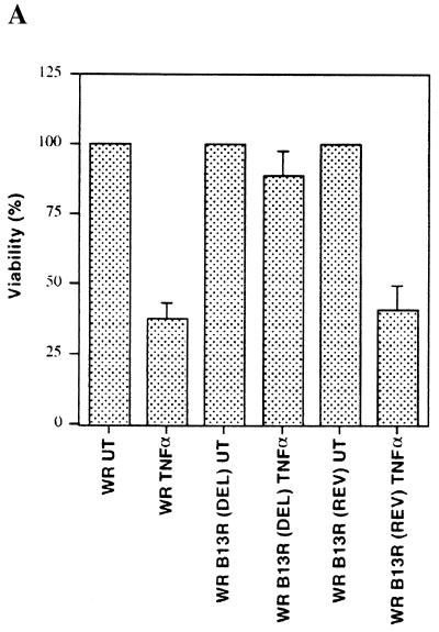
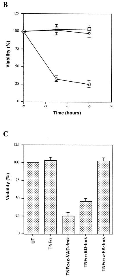
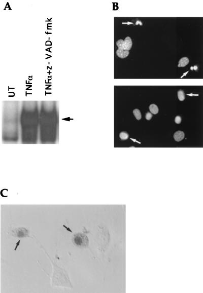
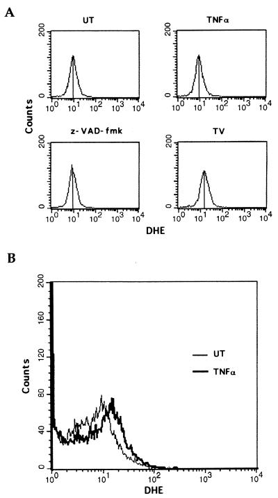
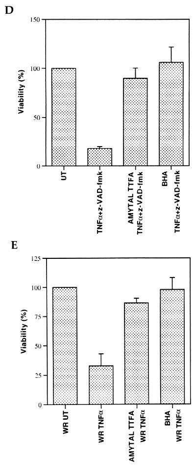
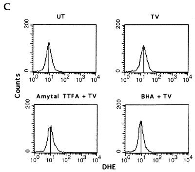
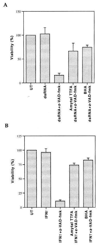
Induction of apoptotic cell death generally requires the participation of cysteine proteases belonging to the caspase family. However, and similar to most cell types, mouse fibroblasts are normally resistant to tumor necrosis factor alpha (TNF-alpha)-induced apoptosis. Surprisingly, TNF-alpha treatment of vaccinia virus-infected mouse fibroblasts resulted in necrotic-like cell death, which was significantly reduced in cells infected with a vaccinia virus mutant lacking the caspase inhibitor B13R. Furthermore, TNF-alpha also induced necrotic-like cell death of fibroblasts in the presence of peptidyl caspase inhibitors. In both cases, necrosis was accompanied by generation of superoxide species. Caspase inhibitors also sensitized fibroblasts to killing by double-stranded RNA and gamma interferon. In all cases, cell death was efficiently blocked by antioxidants or mitochondrial respiratory chain inhibitors. These results define a new mitochondrion-dependent mechanism which may be important in the killing of cells infected with viruses encoding caspase inhibitors.


Figures

FIG. 1
FIG. 1
Caspase inhibitors sensitize mouse fibroblasts to TNF-α-mediated cytotoxicity. (A) 3T3 fibroblasts were infected with either WR strain wild-type vaccinia virus or B13R deletion (DEL) or B13R revertant (REV) vaccinia virus before being treated with TNF-α (1 ng/ml) for 3 h. Viable cells after treatment are shown as a percentage of viable untreated cells (UT). (B) 3T3 fibroblasts were treated with TNF-α (□), z-VAD-fmk (◊), or TNF-α plus z-VAD-fmk (○) for 3 or 6 h. (C) 3T3 fibroblasts were treated with TNF-α alone or with z-VAD-fmk, BD-fmk, or z-FA-fmk for 6 h.
FIG. 1
FIG. 1
Caspase inhibitors sensitize mouse fibroblasts to TNF-α-mediated cytotoxicity. (A) 3T3 fibroblasts were infected with either WR strain wild-type vaccinia virus or B13R deletion (DEL) or B13R revertant (REV) vaccinia virus before being treated with TNF-α (1 ng/ml) for 3 h. Viable cells after treatment are shown as a percentage of viable untreated cells (UT). (B) 3T3 fibroblasts were treated with TNF-α (□), z-VAD-fmk (◊), or TNF-α plus z-VAD-fmk (○) for 3 or 6 h. (C) 3T3 fibroblasts were treated with TNF-α alone or with z-VAD-fmk, BD-fmk, or z-FA-fmk for 6 h.
FIG. 2
FIG. 2
TNF-α induces apoptosis in the presence of macromolecule synthesis inhibitors. (A) Mouse 3T3 fibroblasts were treated with TNF-α plus actinomycin D (2 μg/ml) for 3 h, after which trypan blue was added. Apoptotic cells are indicated by arrows. (B) Cell lysates from untreated (UT) or TNF-α–actinomycin D-treated (TA) cells were made in the presence of biot-VAD-fmk (1 μM), biot-VAD-fmk and z-VAD-fmk, or biot-VAD-fmk and z-FA-fmk, and lysates (20 μg of protein) were examined by a biotin-avidin affinity blot. The activated putative caspase P1 is indicated. (C) Whole-cell lysates from cells treated as in panel B were analyzed by a Western blot assay with a PARP-specific antibody. The 116-kDa precursor and the 85-kDa processed form are indicated. (D) Mock-infected or vaccinia virus-infected 3T3 fibroblasts were left untreated (UT) or treated with TNF-α plus cycloheximide (TC) for 3 h, after which P1 was detected as in panel B. (E) 3T3 fibroblasts were treated with TNF-α plus actinomycin D (□), z-VAD-fmk (◊), or TNF-α plus actinomycin D and z-VAD-fmk (○) for 3 or 6 h. Viable cells remaining after treatment are shown as a percentage of viable untreated cells. DEL, B13R deletion mutant; REV, B13R revertant.
FIG. 3
FIG. 3
TNF-α–caspase inhibitor treatment induces necrotic-like cell death. (A) 3T3 fibroblasts were pretreated with z-VAD-fmk for 1 h or left untreated (UT) before the addition of TNF-α for 1 h. Nuclear extracts were made to test NF-κB activation. The arrow indicates the mobility of the NF-κB–DNA complex. (B) TNF-α–actinomycin D-treated (top) or TNF-α–z-VAD-fmk-treated (bottom) cells were stained with DAPI. Nuclear morphology is indicated by arrows. (C) 3T3 fibroblasts were treated with TNF-α plus z-VAD-fmk for 3 h, and trypan blue was added. Arrows indicate typical necrotic cells.
FIG. 4
FIG. 4
TNF-α–caspase inhibitor treatment induces the production of superoxides. (A) 3T3 fibroblasts were either left untreated (UT) or treated with TNF-α, z-VAD-fmk, or TNF-α plus z-VAD-fmk (TV) for 3 h. Intracellular superoxide levels were detected by DHE binding. (B) WR strain wild-type vaccinia virus-infected cells were either left untreated (UT) or treated with TNF-α (1 ng/ml) for 3 h, after which intracellular superoxide levels were detected by DHE binding. (C) 3T3 fibroblasts were left untreated (UT) or treated with TNF-α plus z-VAD-fmk (TV) for 3 h in the absence or presence of mitochondrial complex I and II inhibitors (amytal and TTFA) or the antioxidant BHA. Superoxide levels were detected as in panel A. (D) 3T3 fibroblasts were treated with TNF-α plus z-VAD-fmk for 6 h in the absence or presence of amytal and TTFA or BHA. Cell viability is shown as a percentage of untreated cells (UT). (E) 3T3 fibroblasts were infected with WR strain wild-type vaccinia virus. They were then treated with TNF-α (1 ng/ml) in the absence or presence of amytal and TTFA or BHA for 3 h.
FIG. 4
FIG. 4
TNF-α–caspase inhibitor treatment induces the production of superoxides. (A) 3T3 fibroblasts were either left untreated (UT) or treated with TNF-α, z-VAD-fmk, or TNF-α plus z-VAD-fmk (TV) for 3 h. Intracellular superoxide levels were detected by DHE binding. (B) WR strain wild-type vaccinia virus-infected cells were either left untreated (UT) or treated with TNF-α (1 ng/ml) for 3 h, after which intracellular superoxide levels were detected by DHE binding. (C) 3T3 fibroblasts were left untreated (UT) or treated with TNF-α plus z-VAD-fmk (TV) for 3 h in the absence or presence of mitochondrial complex I and II inhibitors (amytal and TTFA) or the antioxidant BHA. Superoxide levels were detected as in panel A. (D) 3T3 fibroblasts were treated with TNF-α plus z-VAD-fmk for 6 h in the absence or presence of amytal and TTFA or BHA. Cell viability is shown as a percentage of untreated cells (UT). (E) 3T3 fibroblasts were infected with WR strain wild-type vaccinia virus. They were then treated with TNF-α (1 ng/ml) in the absence or presence of amytal and TTFA or BHA for 3 h.
FIG. 5
FIG. 5
Caspase inhibitors sensitize mouse fibroblasts to dsRNA-induced or IFN-γ-induced cytotoxicity. (A) 3T3 fibroblasts were left untreated (UT) or treated with dsRNA or with dsRNA plus z-VAD-fmk for 6 h in the absence or presence of mitochondrial complex I and II inhibitors (amytal and TTFA) or the antioxidant BHA. Viable cells after treatment are shown as a percentage of viable untreated cells. (B) 3T3 fibroblasts were left untreated (UT) or treated with IFN-γ or with IFN-γ plus z-VAD-fmk for 6 h in the absence or presence of amytal and TTFA or BHA.

References

    1. Ashkenazi A, Dixit V M. Death receptors: signaling and modulation. Science. 1998;281:1305–1308. - PubMed
    1. Beg A A, Baltimore D. An essential role for NF-kappaB in preventing TNF-alpha-induced cell death. Science. 1996;274:782–784. - PubMed
    1. Beg A A, Sha W C, Bronson R T, Ghosh S, Baltimore D. Embryonic lethality and liver degeneration in mice lacking the RelA component of NF-κB. Nature. 1995;376:167–170. - PubMed
    1. Boehm U, Klamp T, Groot M, Howard J C. Cellular responses to interferon-gamma. Annu Rev Immunol. 1997;15:749–795. - PubMed
    1. Carswell E A, Old L J, Kassel R L, Green S, Fiore N, Williamson B. An endotoxin-induced serum factor that causes necrosis of tumors. Proc Natl Acad Sci USA. 1975;72:3666–3670. - PMC - PubMed

Publication types

LinkOut - more resources