. 2000 Aug;74(16):7518-28.
doi: 10.1128/jvi.74.16.7518-7528.2000.

Effects of deletion or stringent repression of the H3L envelope gene on vaccinia virus replication

Affiliations

Effects of deletion or stringent repression of the H3L envelope gene on vaccinia virus replication

F G da Fonseca et al. J Virol. 2000 Aug.

Abstract

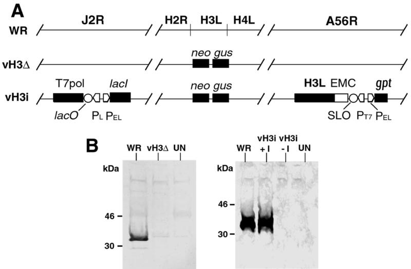
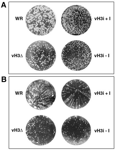
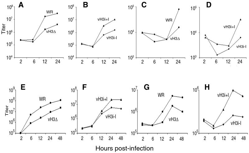
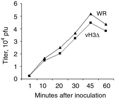
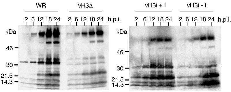
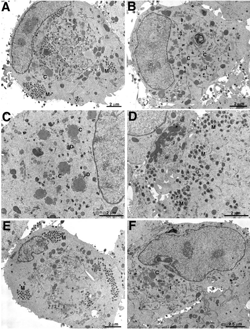
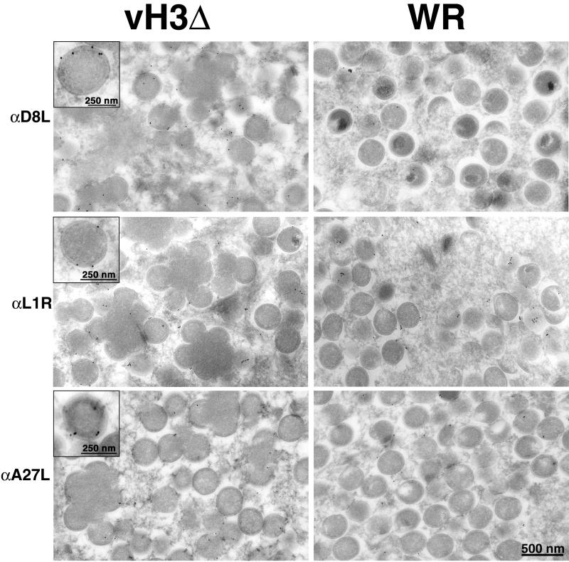
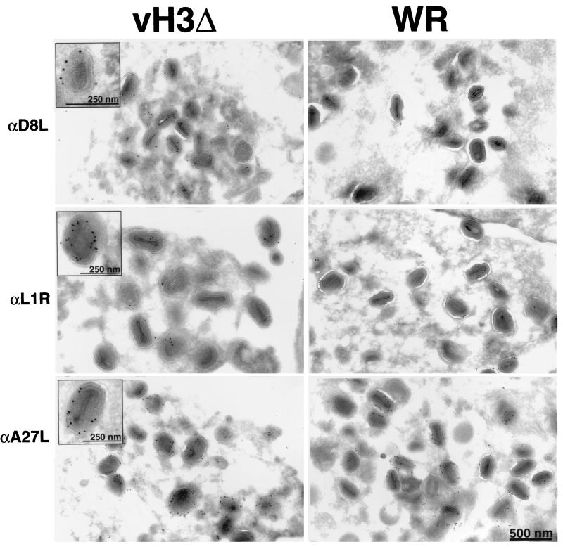
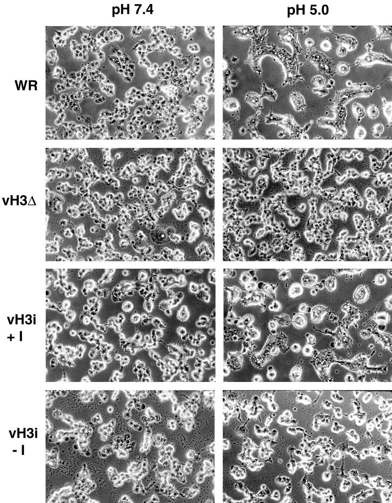
The C-terminal membrane anchor protein encoded by the H3L open reading frame of vaccinia virus is located on the surfaces of intracellular mature virions. To investigate the role of the H3L protein, we constructed deletion (vH3Delta) and inducible (vH3i) null mutants. The H3L protein was not detected in lysates of cells infected with vH3Delta or vH3i in the absence of inducer. Under these conditions, plaques were small and round instead of large and comet shaped, indicative of decreased virus replication or cell-to-cell spread. The mutant phenotype was correlated with reduced yields of infectious intra- and extracellular virus in one-step growth experiments. The defect in vH3i replication could not be attributed to a role of the H3L protein in virus binding, internalization, or any event prior to late gene expression. Electron microscopic examination of cells infected with vH3Delta or vH3i in the absence of inducer revealed that virion assembly was impaired, resulting in a high ratio of immature to mature virus forms with an accumulation of crescent membranes adjacent to granular material and DNA crystalloids. The absence of the H3L protein did not impair the membrane localization of virion surface proteins encoded by the A27L, D8L, and L1R genes. The wrapping of virions and actin tail formation were not specifically blocked, but there was an apparent defect in low-pH-mediated syncytium formation that could be attributed to decreased virus particle production. The phenotypes of the H3L deletion and repression mutants were identical to each other but differed from those produced by null mutations of genes encoding other vaccinia virus membrane components.


Figures

FIG. 1
FIG. 1
Representation of wild-type and mutant genomes. (A) Genomes of wild-type and recombinant viruses. Shown are genome segments containing the following ORFs: J2R (encoding thymidine kinase) H2R, H3L, H4L, and A56R (encoding hemagglutinin). In vH3Δ, the H3L ORF is replaced by the neo and gus genes, which are regulated by vaccinia virus promoters and which provide antibiotic selection and positive identification of plaques. In vH3i, (i) the H3L ORF is replaced by the neo and gus genes regulated by vaccinia virus promoters, (ii) the bacteriophage T7 RNA polymerase ORF regulated by the E. coli lac operator (lacO) and the vaccinia virus P11 late promoter (PL) and the E. coli lac repressor (lacI) regulated by the vaccinia virus P7.5 early/late promoter (PEL) are inserted into the J2R ORF, and (iii) the H3L ORF preceded by the encephalomyocarditis virus untranslated leader sequence (EMC) providing cap-independent translation, a modified lacO (SLO), and a bacteriophage T7 promoter (PT7) as well as the E. coli gpt gene regulated by a vaccinia virus promoter for antibiotic selection are inserted into the A56R ORF. (B) Expression of the H3L ORF. BS-C-1 cells that were uninfected (UN) or infected with WR, vH3Δ, or vH3i in the presence (+I) or absence (−I) of IPTG were harvested after 24 h and the lysates were analyzed by SDS-PAGE and Western blotting with antiserum to a peptide encoded by the H3L ORF. The H3L protein was detected by chemiluminescence. The positions and masses of marker proteins are indicated at the left.
FIG. 2
FIG. 2
Plaque phenotypes of mutant viruses. (A) BS-C-1 cell monolayers were inoculated with WR, vH3Δ, or vH3i in the presence (+I) or absence (−I) of IPTG and stained with crystal violet after 48 h. (B) Same as panel A except that RK13 cells were used.
FIG. 3
FIG. 3
One-step growth curves. BS-C-1 cells (A to D) and RK13 cells (E to H) were infected with purified WR, vH3Δ, or vH3i in the presence (+I) or absence (−I) of IPTG as indicated. At 2, 6, 12, 24, and 48 h, the medium (C, D, G, and H) and cells (A, B, E, and F) were harvested separately and infectious virus titers were determined by plaque assay.
FIG. 4
FIG. 4
Binding of infectious WR and vH3Δ to cells. Replicate wells of chilled BS-C-1 cells were inoculated with 1 PFU of unpurified WR or vH3Δ per cell and maintained on ice. At 1, 10, 20, 30, 45, and 60 min, cells were washed with cold PBS, harvested, dispersed, and plated on fresh BS-C-1 monolayers at 37°C. After 48 h, the cells were stained with crystal violet and the plaques were enumerated.
FIG. 5
FIG. 5
Synthesis of viral proteins. BS-C-1 cells were infected with WR, vH3Δ, or vH3i in the presence (+I) or absence (−I) of IPTG. At 2, 6, 12, 18, and 24 h after infection, cells were harvested and total proteins were subjected to SDS-PAGE. The resolved proteins were transferred to membranes, probed with antiserum made in rabbits infected with purified vaccinia virus, and detected by chemiluminescence. The positions and masses of marker proteins are shown on the left.
FIG. 6
FIG. 6
Electron microscopy of ultrathin sections of infected cells. RK13 cells were infected with WR (A), vH3Δ (B to D), or vH3i in the presence (E) or absence (F) of IPTG and incubated for 23 h. Abbreviations: C, crescents; I, IV; M, mature virions; D, DNA crystalloids. Magnification is indicated by bars.
FIG. 7
FIG. 7
Detection of D8L, L1R, and A27L proteins on membranes of IV formed in cells infected with vH3Δ or WR. Infected cells were cryosectioned and incubated with antibody (α) to D8L, L1R, or A27L followed by protein A-gold. Insets show high magnification of IV selected for good visualization of gold grains overlying the membrane.
FIG. 8
FIG. 8
Detection of D8L, L1R, and A27L proteins on virion membranes of cells infected with vH3Δ or WR. Immunogold labeling was as described in the legend to Fig. 7. Insets show mature virions selected for good visualization of gold grains overlying the membrane.
FIG. 9
FIG. 9
Low-pH-induced fusion of infected cells. BS-C-1 cells were infected with WR, vH3Δ, or vH3i in the presence (+I) or absence (−I) of IPTG. At 12 h after infection, the cells were immersed briefly in buffer at pH 5.0 or 7.4. The medium was replaced, and the incubation continued for an additional 3 h. The cells were photographed with a phase-contrast microscope.

References

    1. Alexander W A, Moss B, Fuerst T R. Regulated expression of foreign genes in vaccinia virus under the control of bacteriophage T7 RNA polymerase and the Escherichia coli lac repressor. J Virol. 1992;66:2934–2942. - PMC - PubMed
    1. Appleyard G, Hapel A J, Boulter E A. An antigenic difference between intracellular and extracellular rabbitpox virus. J Gen Virol. 1971;13:9–17. - PubMed
    1. Blasco R, Moss B. Extracellular vaccinia virus formation and cell-to-cell virus transmission are prevented by deletion of the gene encoding the 37,000-dalton outer envelope protein. J Virol. 1991;65:5910–5920. - PMC - PubMed
    1. Blasco R, Moss B. Role of cell-associated enveloped vaccinia virus in cell-to-cell spread. J Virol. 1992;66:4170–4179. - PMC - PubMed
    1. Boulter E A, Appleyard G. Differences between extracellular and intracellular forms of poxvirus and their implications. Prog Med Virol. 1973;16:86–108. - PubMed

Publication types

MeSH terms

Substances

LinkOut - more resources