Comparative Study
. 2000 Aug;74(16):7578-86.
doi: 10.1128/jvi.74.16.7578-7586.2000.

The structure of cucumber mosaic virus and comparison to cowpea chlorotic mottle virus

Affiliations
Comparative Study

The structure of cucumber mosaic virus and comparison to cowpea chlorotic mottle virus

T J Smith et al. J Virol. 2000 Aug.

Abstract

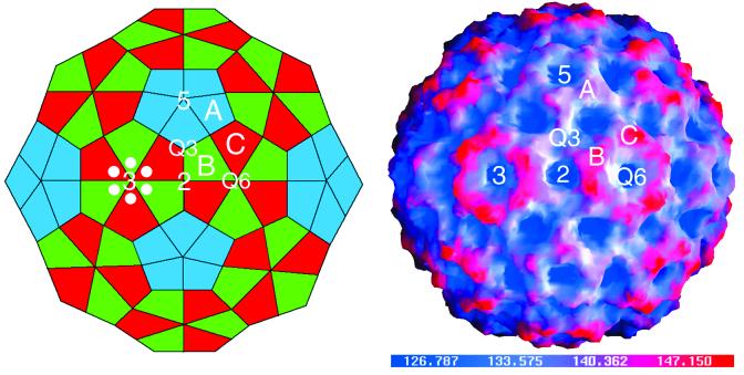
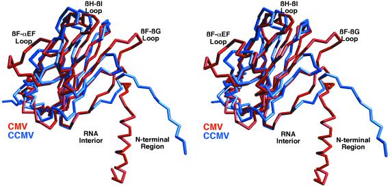
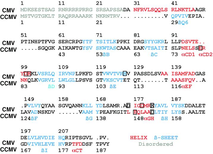
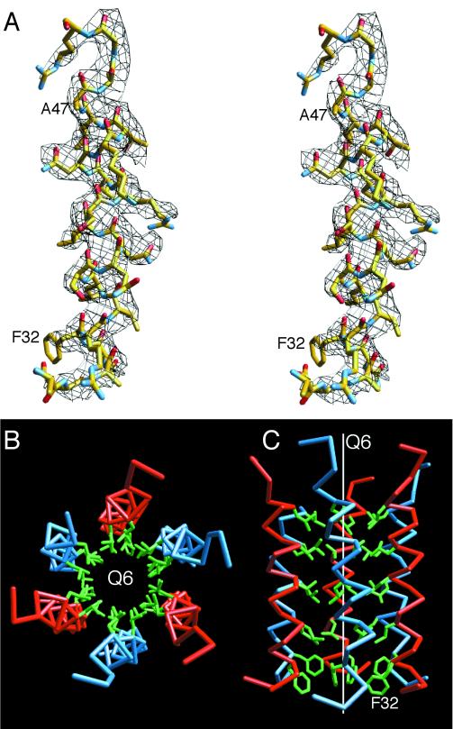
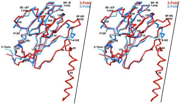
The structure of cucumber mosaic virus (CMV; strain Fny) has been determined to a 3.2-A resolution using X-ray crystallography. Despite the fact that CMV has only 19% capsid protein sequence identity (34% similarity) to cowpea chlorotic mottle virus (CCMV), the core structures of these two members of the Bromoviridae family are highly homologous. As suggested by a previous low-resolution structural study, the 305-A diameter (maximum) of CMV is approximately 12 A larger than that of CCMV. In CCMV, the structures of the A, B, and C subunits are nearly identical except in their N termini. In contrast, the structures of two loops in subunit A of CMV differ from those in B and C. These loops are 6 and 7 residues longer than the analogous regions in CCMV. Unlike that of CCMV, the capsid of CMV does not undergo swelling at pH 7.0 and is stable at pH 9.0. This may be partly due to the fact that the N termini of the B and C subunits form a unique bundle of six amphipathic helices oriented down into the virion core at the threefold axes. In addition, while CCMV has a cluster of aspartic acid residues at the quasi-threefold axis that are proposed to bind metal in a pH-dependent manner, this cluster is replaced by complementing acids and bases in CMV. Finally, this structure clearly demonstrates that the residues important for aphid transmission lie at the outermost portion of the betaH-betaI loop and yields details of the portions of the virus that are hypothesized to mediate binding to aphid mouthparts.


Figures

FIG. 1
FIG. 1
Schematic representation of the T=3, truncated icosahedron (left) and a surface representation of the CMV capsid colored according to radial distance (right). In the schematic, the labeled A, B, and C subunits are those that are in the general orientation used for the following diagrams. The subunits used to represent the icosahedral asymmetric unit were chosen to demonstrate the quasi-sixfold axis and are not related by a quasi-threefold axis. In both images the icosahedral threefold (quasi-sixfold [Q6]), fivefold, twofold, and the quasi-threefold (Q3) axes are labeled. In the schematic, six white circles are positioned around one of the quasi-sixfold axes to approximate the location of the hexameric bundle of N-terminal helices described in the following figures.
FIG. 2
FIG. 2
Comparison of C subunits of CMV and CCMV. The C-α backbone of CMV is shown in red, and that of CCMV is shown in blue. The program MolView was used for this alignment, and 91 residues yielded a root-mean-square deviation of 1.3 Å. Some of the key areas of differences are labeled.
FIG. 3
FIG. 3
Sequence homology of CMV and CCMV based on structural alignments. The gray regions represent disordered regions, the red regions are helices, and the blue regions are β-strands. The nomenclature used for secondary elements is the same as that used for CCMV. The boxed amino acids are those involved in the subunit contacts about the quasi-threefold axes.
FIG. 4
FIG. 4
Amphipathic hexameric helical bundles found at the quasi-sixfold axes. (A) A stereo view of the electron density at 1 ς and the fitted model at the N termini of the C subunits are shown. The model is colored according to atom type. (B and C) Six helices about the quasi-sixfold axis (Q6). The C-α backbones for the B and C subunits are shown in blue and red, respectively. The side chains of the inner, hydrophobic residues are shown in green. All the residues shown in green are leucine except for the most N-terminal residue, which is a phenylalanine. The view from the capsid toward the RNA interior in panel B is parallel to the icosahedral axis, whereas the view in panel C is perpendicular to the quasi-sixfold axis.
FIG. 5
FIG. 5
Comparison of the CMV A- and C-subunit structures. The C-α backbones of the A and C subunits are shown in blue and red, respectively. The RNA interior is toward the bottom of the diagram. The approximate locations of the threefold axis (for the C subunit) and the fivefold axis (for the A subunit) are represented by the black lines. C-Term, C terminus.
FIG. 6
FIG. 6
Subunit interactions about the quasi-threefold axes. For clarity, only the interactions between two of three subunits are shown. The C-α backbone of an A subunit is shown in black, and that of a C subunit is shown in purple. The side chains of the residues at this interface are colored according to atom type: nitrogen atoms are blue, carbon atoms are yellow, and oxygen atoms are red. In CCMV, this same interface is entirely composed of acidic residues that are proposed to interact via a divalent cation.
FIG. 7
FIG. 7
Structure of an external loop involved in aphid transmission. (A) van der Waals surface of a portion of the CMV capsid, with the negatively and positively charged electrostatic fields being shown in red and blue, respectively, created with the program GRASP (15). Arrows denote the locations of the loops described below. Note that the only negatively charged patch on the entire capsid surface is about the βH-βI loop. (B) Distances between the residues involved in this negatively charged patch. The atoms are colored according to atom type as defined in the legend to Fig. 5. The side chains in the area are very close to each other and may be indicative of a counterbalancing cation. (C) Same region and view as those shown in panel B, with the electron density contoured at 1 ς, represented by black lines. Note the patch of density between D118, S119, E198, and D192, which may represent a bound, divalent cation. The distances between the center of this patch of density and the oxygen atoms are between 2.3 and 3.0 Å. Upon deprotonation at neutral pH, these distances may decrease to those of typical oxygen-calcium contacts (∼2.3 Å). Q3 and Q6, quasi-three- and quasi-sixfold axes, respectively.
FIG. 8
FIG. 8
Locations of some of the mutations that affect aphid transmission and movement within the plant. The C-α backbones of one of the A, B, and C subunits are shown in blue, green, and red, respectively. The surrounding icosahedrally related subunits are shown in gray. The nearest five- and threefold (quasi-sixfold [Q6]) axes are labeled. The positions of the various mutation sites are represented by colored spheres. P129, represented by yellow balls, appears to be involved in aphid transmission and host symptomalogy. S129F can be compensated for by mutations that lie on αEF (residues 138, 144, and 147), represented by black balls. Mutations at A162 (cyan balls) affect aphid transmission, perhaps by decreasing capsid stability. The destabilizing effects of deleting residues 15 to 40 can be partially circumvented by the mutations at residues 81, 166, and 173, denoted by mauve balls.

References

    1. Abad-Zapatero C, Abdel-Meguid S S, Johnson J E, Leslie A G W, Rayment I, Rossmann M G, Suck D, Tsukihara T. Structure of southern bean mosaic virus at 2.8Å resolution. Nature (London) 1980;286:33–39. - PubMed
    1. Bailey S. The CCP4 suite: programs for protein crystallography. Acta Crystallogr Sect D. 1994;50:760–763. - PubMed
    1. Bancroft J B, Hills G J, Markham R. A study of the self-assembly process in a small spherical virus. Formation of organized structures from protein subunits in vitro. Virology. 1967;31:354–379. - PubMed
    1. Collaborative Computational Project n. The CCP4 suite: programs for protein crystallography. Acta Crystallogr Sect D. 1994;50:760–763. - PubMed
    1. Gray S M. Plant virus proteins involved in natural vector transmission. Trends Microbiol. 1996;4:259–264. - PMC - PubMed

Publication types