. 2000 Jul 15;28(14):2643-50.
doi: 10.1093/nar/28.14.2643.

Common fold in helix-hairpin-helix proteins

Affiliations

Common fold in helix-hairpin-helix proteins

X Shao et al. Nucleic Acids Res. .

Abstract

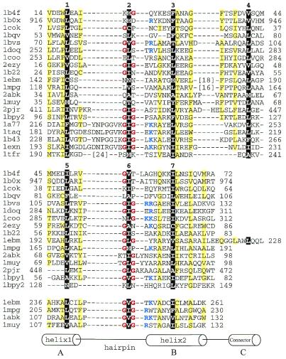
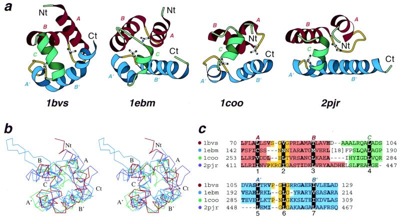
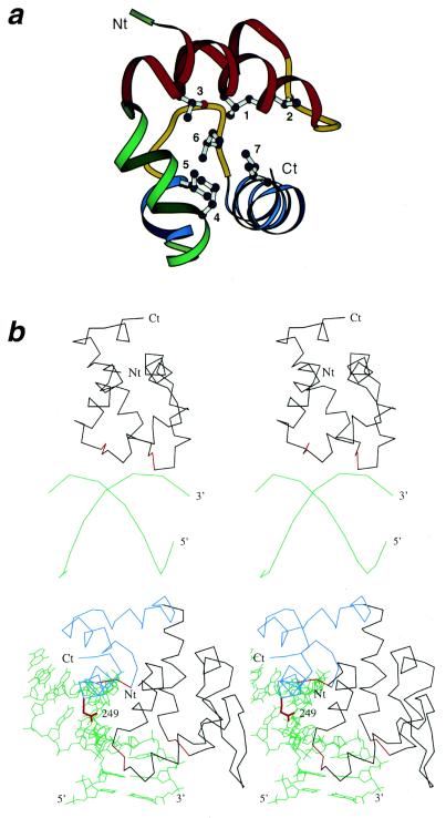
Helix-hairpin-helix (HhH) is a widespread motif involved in non-sequence-specific DNA binding. The majority of HhH motifs function as DNA-binding modules, however, some of them are used to mediate protein-protein interactions or have acquired enzymatic activity by incorporating catalytic residues (DNA glycosylases). From sequence and structural analysis of HhH-containing proteins we conclude that most HhH motifs are integrated as a part of a five-helical domain, termed (HhH)(2) domain here. It typically consists of two consecutive HhH motifs that are linked by a connector helix and displays pseudo-2-fold symmetry. (HhH)(2) domains show clear structural integrity and a conserved hydrophobic core composed of seven residues, one residue from each alpha-helix and each hairpin, and deserves recognition as a distinct protein fold. In addition to known HhH in the structures of RuvA, RadA, MutY and DNA-polymerases, we have detected new HhH motifs in sterile alpha motif and barrier-to-autointegration factor domains, the alpha-subunit of Escherichia coli RNA-polymerase, DNA-helicase PcrA and DNA glycosylases. Statistically significant sequence similarity of HhH motifs and pronounced structural conservation argue for homology between (HhH)(2) domains in different protein families. Our analysis helps to clarify how non-symmetric protein motifs bind to the double helix of DNA through the formation of a pseudo-2-fold symmetric (HhH)(2) functional unit.


Figures

Figure 1
Figure 1
Structure-based sequence alignment of HhH proteins. For each sequence, PDB entry name and starting and ending residue numbers are given. Protein name, chain name (if any) and gene identifier (gi) number for each entry are: 1b4f, human EphB2 receptor, chain A, 4558093; 1b0x, mouse EphA4 receptor tyrosine kinase, chain A, 4929864; 1cok, human p73 C-terminal domain, chain A, 5822025; 1bqv, mouse Ets-a transcription factor pointed domain, 3891925; 1bvs, Mycobacterium leprae DNA-helicase RuvA middle domain, chain A, 3660156; 1doq, T.thermophilus RNA-polymerase α-subunit C-terminal domain, chain A, 6730428; 1coo, E.coli RNA-polymerase α-subunit C-terminal domain, 1421046; 2ezy, human BAF, chain A, 4389121; 1b22, human DNA repair protein Rad51 N-terminal domain, chain A, 6730074; 1ebm, human 8-oxoguanine glycosylase central domain, chain A, 2078294; 1mpg, E.coli 3-methyladenine DNA glycosylase II 2 C-terminal domains, chain A, 2914353; 2abk, E.coli endonuclease III, 1311214; 1muy, E.coli MutY catalytic domain, chain A, 5822134; 2pjr, Bacillus stearothermophilus DNA-helicase PcrA insertion domain, chain A, 4930184; 1bpy1 and 1bpy2, human DNA-polymerase beta N-terminal (8 kDa) domain and ‘fingers’ domain, respectively, chain A, 2392200; 1a77, Methanococcus jannaschii Flap endonuclease-1, 5821778; 1taq, Thermus aquaticus DNA-polymerase Taq 5′ to 3′ exonuclease domain, 1942938; 1b43, Pyrococcus furiosus Fen-1 nuclease, chain A, 6980604; 1exn, bacteriophage T5 5′-exonuclease, chain A, 2392326; 1tfr, bacteriophage T4 RNase H, 1943457. The first HhH motifs are aligned in the top panel. For proteins with more than one HhH motif, the second and the third motifs are aligned in the middle and the bottom panels, respectively. All three panels are also aligned with each other. Positions of hydrophobic core residues are highlighted in black and are labeled with numbers corresponding to those in Figure 2c. Residues in the third HhH motif are not numbered. Glycines in the signature sequence GhG (h is a hydrophobic residue) of the hairpin regions are in red and positively charged residues following the signature are in blue. Positions with mostly uncharged residues are shaded in yellow. Numbers in brackets indicate the number of omitted residues in the sequence.
Figure 2
Figure 2
Structural comparisons of divergent (HhH)2 domains. (a) Structural diagrams of DNA-helicase RuvA middle domain (1bvs, chain A, residues 63–134), 8-oxoguanine glycosylase central domain (1ebm, chain A, 135–221), C-terminal domain of RNA-polymerase α-subunit (1coo, 253–296) and DNA-helicase PcrA insertion domain (2pjr, chain A, 405–478), showing the HhH motifs from each protein. For each protein, N- and C-termini are labeled with Nt and Ct, respectively. The helices in the first HhH motif are labeled with A and B, and are in red. The corresponding helices in the second HhH motif are labeled with A′ and B′ and are in blue. The hairpin regions of both motifs are in yellow. Side-chains of central hydrophobic residues in hairpins are shown using ball-and-stick representation. The helices connecting the two HhH motifs are labeled with C and are in green. The ribbon diagrams were rendered by Bobscript (28), a modified version of Molscript (29). (b) Stereo diagram of superimposed Cα traces of DNA-helicase RuvA subunit (red), 8-oxoguanine glycosylase (blue), C-terminal domain of RNA-polymerase α-subunit (green) and DNA-helicase PcrA (purple). Superpositions were made using InsightII package (Molecular Simulations Inc.). Labels match those described in (a). (c) Structure-based sequence alignment of HhH motif regions of the four illustrated protein domains. For each sequence the PDB entry name and starting and ending residue numbers are given. The dot color scheme (in front of each PDB entry) matches those in (b). Color shading and helix labels correspond to those in (a). The two HhH motifs (upper and lower panels) are aligned with each other. Sites of conserved core hydrophobic residues are highlighted in black, and are labeled 1, 2 and 3 in the first HhH motif, 5, 6 and 7 in the second motif, and 4 in the connector helix C (also see Fig. 1). Additional conserved hydrophobic residues are shown in bold.
Figure 3
Figure 3
Conserved hydrophobic core residues in the (HhH)2 proteins and DNA binding by HhH proteins. (a) Ribbon diagram of DNA-helicase RuvA middle domain (1bvs, chain A, residues 63–134) showing its hydrophobic core. The coloring and termini labeling scheme follows that in Figure 1a. Conserved core hydrophobic residues L74, V80, T88, L109, L115 and I123 are shown using ball-and-stick representation, and are labeled with numbers 1–7 corresponding to those in Figures 1c and 2. (b) Stereo diagrams showing interaction of (HhH)2 domains and DNA. Upper panel: E.coli DNA-helicase RuvA middle domain (1bdx, chain A, residues 65–142) with bound DNA at a Holliday junction (alpha carbons and phosphate atoms only). Lower panel: human 8-oxoguanine glycosylase (1ebm, 135–262) with bound DNA. DNA chains are in green and termini are labeled with 5′ and 3′. Protein Cα traces are in black, except that the third (catalytic) HhH motif in human 8-oxoguanine glycosylase is in blue, and the signature sequences GhG of the hairpin regions are in red. N- and C-termini are labeled with Nt and Ct, respectively. Side chain of residue 249 (Lys, mutated to Gln in the structure) in human 8-oxoguanine glycosylase, which is involved in the lyase activity of the protein, is shown in red.

References

    1. Bateman A., Birney,E., Durbin,R., Eddy,S.R., Finn,R.D. and Sonnhammer,E.L. (1999) Nucleic Acids Res., 27, 260–262. - PMC - PubMed
    1. McClure M.A., Smith,C. and Elton,P. (1996) ISMB, 4, 155–164. - PubMed
    1. Hughey R. and Krogh,A. (1996) Comput. Appl. Biosci., 12, 95–107. - PubMed
    1. Altschul S.F., Madden,T.L., Schaffer,A.A., Zhang,J., Zhang,Z., Miller,W. and Lipman,D.J. (1997) Nucleic Acids Res., 25, 3389–3402. - PMC - PubMed
    1. Altschul S.F. and Koonin,E.V. (1998) Trends Biochem. Sci., 23, 444–447. - PubMed

MeSH terms