. 2000 Jul 15;28(14):2741-51.
doi: 10.1093/nar/28.14.2741.

PTIP, a novel BRCT domain-containing protein interacts with Pax2 and is associated with active chromatin

Affiliations

PTIP, a novel BRCT domain-containing protein interacts with Pax2 and is associated with active chromatin

M S Lechner et al. Nucleic Acids Res. .

Abstract

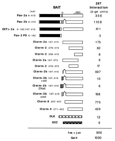
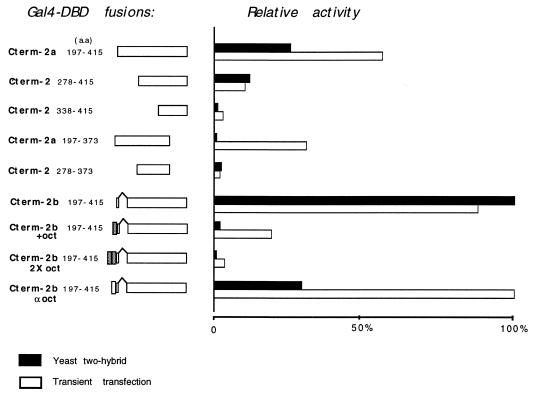
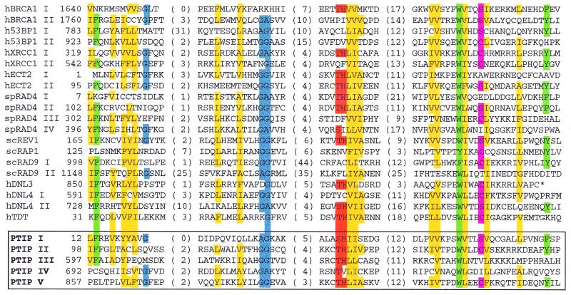
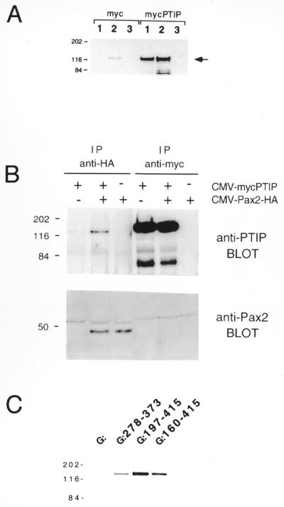
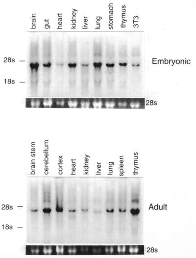
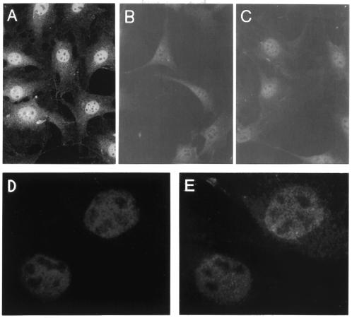
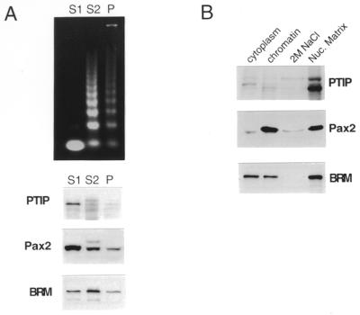
The Pax gene family encodes transcription factors essential for organ and tissue development in higher eukaryotes. Pax proteins are modular with an N-terminal DNA binding domain, a C-terminal transcription activation domain, and a transcription repression domain called the octapeptide. How these domains interact with the cellular machinery remains unclear. In this report, we describe the isolation and characterization of a novel gene and its encoded protein, PTIP, which binds to the activation domain of Pax2 and other Pax proteins. PTIP binds to Pax2 in vitro, in the yeast two-hybrid assay and in tissue culture cells. The binding of PTIP to Pax2 is inhibited by the octapeptide repression domain. The PTIP protein contains five BRCT domains, first identified in BRCA1 and other nuclear proteins involved in DNA repair/recombination or cell cycle control. Pax2 and PTIP co-localize in the cell nucleus to actively expressed chromatin and the nuclear matrix fraction. For the first time, these results point to a link between Pax transcription factors and active chromatin.


Figures

Figure 1
Figure 1
Interaction between clone 287 (PTIP) and Pax2 in the yeast two-hybrid system. Yeast strain MaV103 was co-transformed with expression plasmids for clone 287 fused to the GAD and Pax2 fused to the GDBD. The portions of Pax2 (or Pax6 and Pax8) encoded by the plasmids are indicated schematically and by amino acid number of the full-length proteins. The paired domain is indicated by a black box, the octapeptide by a shaded box and the region rich in proline, serine, threonine and tyrosine residues by PSTY. Control baits were the mixed lineage kinase DLK and netrin receptor DCC fused to the GDBD. Co-transformants were grown in liquid medium and assayed for activation of the lacZ reporter by measuring β-galactosidase activity. At least two independent clones were tested and an average value is given. The background activity of each GDBD–Pax2 fusion alone is subtracted from this value in every case.
Figure 2
Figure 2
Relative transcription activation activity of the Pax2 C-terminal domains and their affinitites for PTIP in the yeast two-hybrid assay. GDBD–Pax2 fusions were tested for activation of a CAT reporter plasmid containing multiple copies of the Gal4 upstream activation sequence (open bars). The same fusion proteins were also analyzed for interaction with PTIP in the two hybrid system (solid bars). The bars indicate the relative average activity for each fusion in the two assays.
Figure 3
Figure 3
Primary amino acid sequence from the conceptual translation of the PTIP cDNA. The polypeptide sequence was deduced from the full-length cDNA which was generated from overlapping yeast and phage clones. The five BRCT domains are underlined and a central glutamine-rich region spans approximately residues 396–577. The complete PTIP sequence is available under GenBank accession no. AF104261.
Figure 4
Figure 4
Alignment of amino acid residues conserved among BRCT domains. Alignment is based upon BLAST analysis of the non-redundant protein sequence database and previously described hydrophobic cluster analysis (36). The first amino acid position of each domain is given as well as the number of intervening residues between conserved blocks. An asterisk indicates a stop codon. Yellow boxes, A, F, G, I, L, V, Y; blue boxes, G, A; green boxes, F, W, Y; pink boxes, S, C; orange boxes, H, S, T. h designates human protein sequences, sc, Saccharomyces cerevisiae and sp, Schizosaccharomyces pombe. The five BRCT domains (I–V) of murine PTIP are outlined by the open box.
Figure 5
Figure 5
Pax2 and PTIP interactions. (A) Specificity of anti-PTIP antibodies. Extracts were prepared from cells transiently transfected with control expression vector (pMYC) or pMYC-PTIP and immunoprecipitated with affinity purified anti-PTIP-1167 (lane 2) or anti-myc monoclonal antibody (lane 1). As a negative control, a chicken anti-mTcf4 antibody was used (lane 3). Immunocomplexes were collected with anti-chicken IgY affinity resin or protein G–Sepharose, washed and separated by SDS–PAGE. Western blots were probed with anti-PTIP-1169. Molecular weight standards are shown to the left (kDa) and the ~130 kDa PTIP protein is indicated by an arrow. Note the endogenous PTIP protein in lane 2, myc panel, and the transfected PTIP protein in lanes 1 and 2, mycPTIP panel. (B) Co-immunoprecipitation of Pax2 and PTIP. NIH 3T3 cells were transiently transfected with expression vectors for epitope-tagged forms of Pax2 and PTIP or a control expression vector (indicated above lanes by + or –). Cell extracts were immunoprecipitated with anti-HA or anti-myc monoclonal antibodies and the immune complexes collected with protein G–Sepharose, washed and separated by SDS–PAGE. Proteins were analyzed by western blotting with polyclonal antisera against either Pax2 or PTIP. Co-immunoprecipitation of PTIP with Pax2 was observed (second lane). Molecular weight standards are shown to the left (kDa). (C) [35S]PTIP from in vitro transcription/translation reactions was incubated with GST–Pax2 fusion proteins. The regions of Pax2b fused to GST are indicated by the amino acids above each lane. After incubation, glutathione–agarose beads were washed and the protein complexes separated by SDS–PAGE. Gels were stained with Coomassie blue to visualize integrity and loading of GST–Pax2 proteins, amplified for fluorography and exposed to film. Experiments for each binding reaction were repeated at least twice and typical results for full-length PTIP are shown.
Figure 6
Figure 6
Northern blot analysis of PTIP mRNA expression. An aliquot of 10 µg of total RNA from various tissues of embryonic and adult mice was separated by denaturing agarose gel electrophoresis, blotted to nylon membrane and probed with a 1 kb EcoRI fragment from the murine PTIP cDNA. 3T3 designates RNA isolated from cultured NIH 3T3 cells. A single PTIP transcript is found with an estimated size of 4000 nt. The position of the 28S and 18S rRNAs are indicated. Variation in RNA loading is shown by the relative intensities of the 28s ribosomal band.
Figure 7
Figure 7
Endogenous PTIP is localized to the nucleus. Affinity purified chicken antisera to murine PTIP were used for indirect immunofluorescent staining of NIH 3T3 cells. (A) Anti-PTIP 1167 antiserum. (B) Preimmune 1167 antiserum. (C) Anti-PTIP 1167 antiserum preincubated with immunizing antigen. (D) Pax2 transformed NIH 3T3 cells stained with anti-Pax2 antibodies. (E) Same cells as (D) stained with anti-PTIP.
Figure 8
Figure 8
Association of PTIP with active chromatin and the nuclear matrix. (A) Micrococcal nuclease digestion of nuclei. (Top) An aliquot of 1 µg DNA extracted from fractions S1, S2 and pellet. Western blots of protein fractions shown below with antibodies against the indicated proteins. (B) Extraction of nuclear matrix. Cells were separated into cytoplasmic fractions, whole chromatin, 2 M NaCl extracted chromatin and insoluble nuclear matrix. Proteins from each fraction were analyzed by western blotting with the indicated antibodies.

References

    1. Dahl E., Koseki,H. and Balling,R. (1997) Bioessays, 19, 755–765. - PubMed
    1. Noll M. (1993) Curr. Opin. Genet. Dev., 3, 595–605. - PubMed
    1. Stuart E.T., Kioussi,C. and Gruss,P. (1993) Annu. Rev. Genet., 27, 219–236. - PubMed
    1. Dressler G.R. and Douglass,E.C. (1992) Proc. Natl Acad. Sci. USA, 89, 1179–1183. - PMC - PubMed
    1. Gnarra J.R. and Dressler,G.R. (1995) Cancer Res., 55, 4092–4098. - PubMed

Publication types

MeSH terms

Associated data