. 2000 Jul 15;28(14):2771-8.
doi: 10.1093/nar/28.14.2771.

Similarities and differences in the conformation of protein-DNA complexes at the U1 and U6 snRNA gene promoters

Affiliations

Similarities and differences in the conformation of protein-DNA complexes at the U1 and U6 snRNA gene promoters

S B Hardin et al. Nucleic Acids Res. .

Abstract

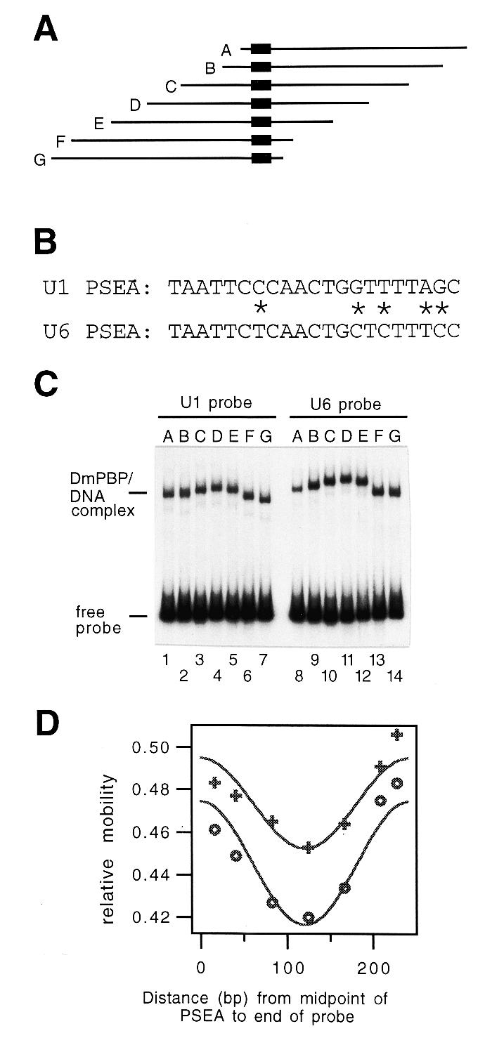
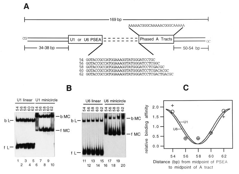
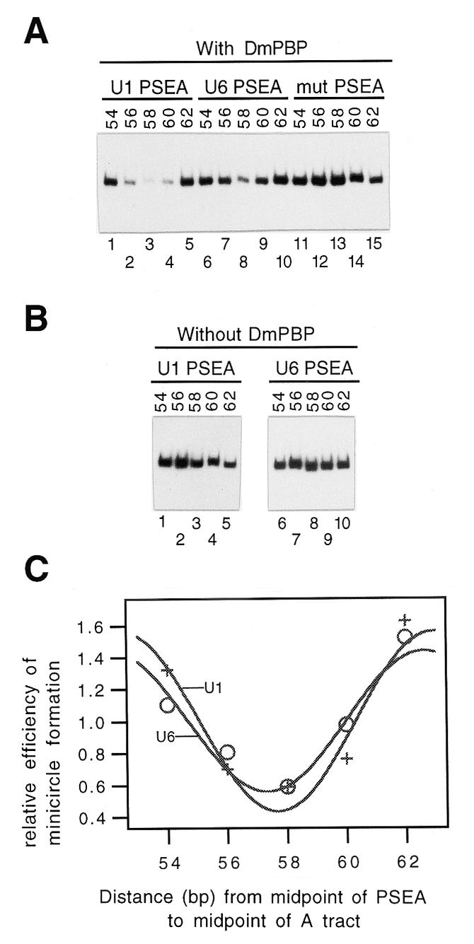
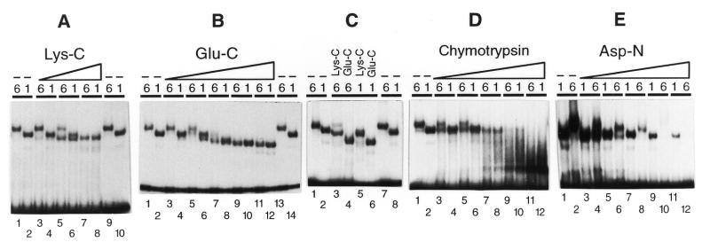
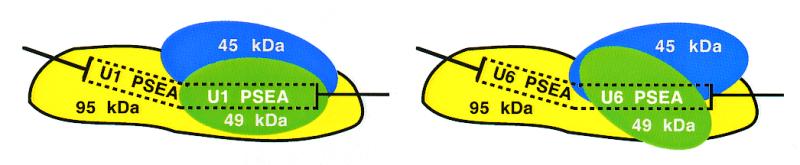
Most small nuclear RNAs (snRNAs) are synthesized by RNA polymerase II, but U6 snRNA is synthesized by RNA polymerase III. In the fruit fly Drosophila melanogaster the RNA polymerase specificity of the snRNA genes is determined by a few nucleotide differences within the proximal sequence element (PSE), a conserved sequence located approximately 40-65 bp upstream of the transcription start site. The PSE is essential for transcription of both RNA polymerase II-transcribed and RNA polymerase III-transcribed snRNA genes and is recognized in Drosophila by a multi-subunit protein factor termed DM:PBP. Previous studies that employed site-specific protein-DNA photocrosslinking indicated that the conformation of the DNA-protein complex is different depending upon whether DM:PBP is bound to a U1 or U6 PSE sequence. These conformational differences of the complex probably represent an early step in determining the selection of the correct RNA polymerase. We have now obtained evidence that DM:PBP modestly bends the DNA upon interacting with the PSE and that the direction of DNA bending is similar for both the U1 and U6 PSEs. Under the assumption that DM:PBP does not significantly twist the DNA, the direction of the bend in both cases is toward the face of the DNA helix contacted by the 45 kDa subunit of DM:PBP. Together with data from partial proteolysis assays, these results indicate that the conformational differences in the complexes of DM:PBP with the U1 and U6 PSEs more likely occur at the protein level rather than at the DNA level.


Figures

Figure 1
Figure 1
Circular permutation assay with DNA fragments that contain a Drosophila U1 or U6 PSEA. (A) Diagram of a set of seven DNA fragments that contain the U1 or U6 PSEA at various positions relative to the ends of the fragments. The location of the PSEA, which is the binding site for DmPBP, within each 242 bp fragment is indicated by the black rectangle. (B) Sequences of the Drosophila U1 and U6 PSEAs with the differences indicated by asterisks. (C) Autoradiogram following native gel electrophoresis of DmPBP–DNA complexes formed with the U1 (lanes 1–7) or the U6 (lanes 8–14) circularly permuted fragments diagrammed in (A). (D) Plot of the relative mobility of DmPBP–DNA complexes versus the position of the PSEA within the fragment. Plus symbols, fragments containing the U1 PSEA; open circles, fragments containing the U6 PSEA.
Figure 2
Figure 2
Binding of DmPBP to pre-bent (minicircle) DNA. (A) The linear fragments that were used to generate the 169 bp minicircles in which the U1 or U6 PSEA would be bent in various directions are diagrammed. The sequences separating the PSEA and phased A tracts in each fragment are shown explicitly, with the distance in base pairs from the midpoint of the PSEA to the midpoint of the A tracts given to the left. (B) Autoradiogram following native gel electrophoresis after incubation of DmPBP with linear fragments or the corresponding minicircular DNA that contained the U1 PSEA (lanes 1–10) or the U6 PSEA (lanes 11–20). bL, bound linear; fL, free linear; bMC, bound minicircle; fMC, free minicircle. (C) Phasing plot of normalized binding affinity versus the distance between the PSEA and the intrinsic bend. Plus symbols, fragments containing the U1 PSEA; open circles, fragments containing the U6 PSEA.
Figure 3
Figure 3
Ligase-catalyzed circularization assays. (A) Following the binding of DmPBP to the DNA fragments diagrammed in Figure 2A, DNA ligase was added and the appearance of circular DNA was monitored by gel electrophoresis following exonuclease digestion. Bands corresponding to 169 bp closed circular DNA are shown. Reactions were performed with DNA fragments that contained a U1 PSEA (lanes 1–5), a U6 PSEA (lanes 6–10) or a mutant PSEA to which DmPBP could not bind (lanes 11–15). (B) Ligation reactions carried out as in (A) but in the absence of the DmPBP fraction. (C) Phasing plot of the normalized efficiency of minicircle formation versus the distance between the PSEA and the intrinsic bend. Plus symbols, fragments containing the U1 PSEA; open circles, fragments containing the U6 PSEA.
Figure 4
Figure 4
Partial proteolysis assays of DmPBP bound to U1 or U6 PSEAs. Labeled DNA fragments that contained the U1 or U6 PSEAs (as indicated by a 1 or 6 above each individual lane) were incubated with DmPBP followed by the addition of increasing amounts of an endoproteinase to partially digest the DmPBP. The free and protein-bound fragments were then separated by native gel electrophoresis and the bands detected by autoradiography. (A) Lanes 3–8, 0.26, 1.3 or 6.4 µg/ml Lys-C; lanes 1, 2, 9 and 10, no proteinase. (B) Lanes 3–12, 0.037, 0.18, 0.91, 4.6 or 23 µg/ml Glu-C; lanes 1, 2, 13 and 14, no proteinase. (C) Lanes 3 and 5, 5.9 µg/ml Lys-C; lanes 4 and 6, 31 µg/ml Glu-C; lanes 1, 2, 7 and 8, no proteinase. (D) Lanes 3–12, 0.17, 0.34, 0.69, 1.4 or 2.8 µg/ml chymotrypsin; lanes 1 and 2, no proteinase. (E) Lanes 3–12, 0.10, 0.31, 0.92, 2.8 or 8.3 µg/ml Asp-N; lanes 1 and 2, no proteinase.
Figure 5
Figure 5
Model of the interaction of DmPBP with Drosophila U1 and U6 gene proximal sequence elements. According to the model, the protein adopts alternative conformations depending upon whether it is bound to a U1 or U6 PSEA sequence. In both cases the DNA is modestly bent upon interacting with DmPBP and the direction of the bend is similar for both the U1 and U6 PSEAs. The figure shows the direction of the DNA bend toward the face of the helix contacted by the 45 kDa subunit under the assumption that DmPBP does not significantly writhe or twist the DNA.

References

    1. Hernandez N. (1992) In McKnight,S.L. and Yamamoto,K.R. (eds), Transcriptional Regulation. Cold Spring Harbor Laboratory Press, Cold Spring Harbor, NY, pp. 281–313.
    1. Lobo S.M. and Hernandez,N.T. (1994) In Conaway,R.C. and Conaway,J.W. (eds), Transcription: Mechanisms and Regulation. Raven Press, New York, NY, pp. 127–159.
    1. Stefanovic B. and Marzluff,W.F. (1992) Mol. Cell. Biol., 12, 650–660. - PMC - PubMed
    1. Wendelburg B.J. and Marzluff,W.F. (1992) Nucleic Acids Res., 20, 3743–3751. - PMC - PubMed
    1. Li J.-M., Parsons,R.A. and Marzluff,W.F. (1994) Mol. Cell. Biol., 14, 2191–2200. - PMC - PubMed

Publication types