. 2000 Aug 1;28(15):2873-81.
doi: 10.1093/nar/28.15.2873.

The endonuclease activity of the yeast Dna2 enzyme is essential in vivo

Affiliations

The endonuclease activity of the yeast Dna2 enzyme is essential in vivo

K H Lee et al. Nucleic Acids Res. .

Abstract

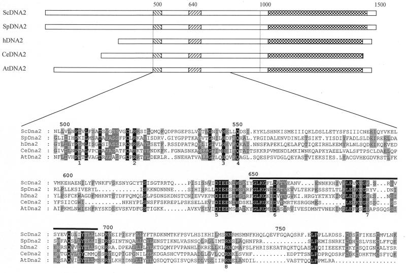
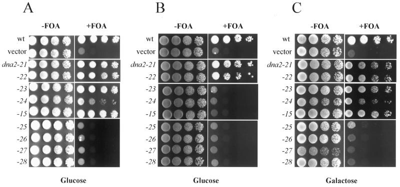
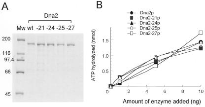
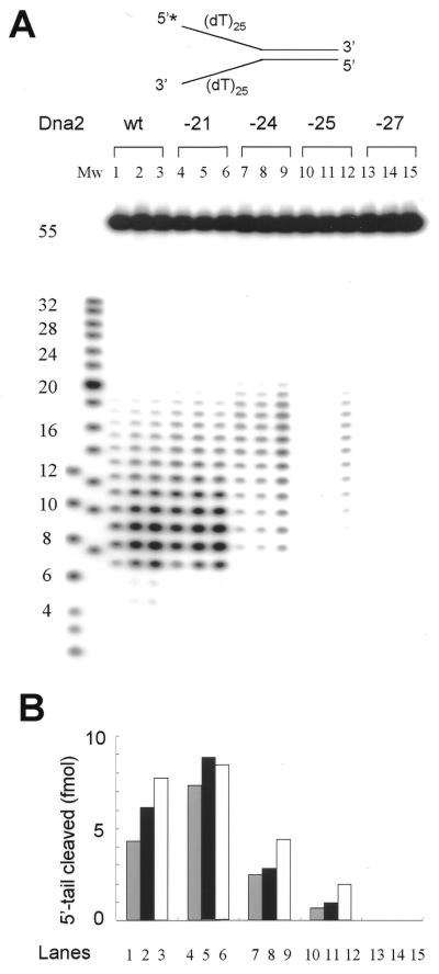
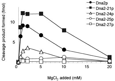
Dna2 is a multifunctional enzyme in yeast that possesses endonuclease activity well suited to remove RNA-DNA primers of Okazaki fragments, raising the question of whether endonuclease activity is essential for in vivo Dna2 function. Systematic site-directed mutations of amino acid residues in Saccharomyces cerevisiae DNA2 conserved in the central region of many eukaryotic DNA2 homologs allowed us to identify mutant dna2 alleles that were divided into three groups based on the viability of the mutant cells: (i) viable; (ii) inviable only when expression was repressed; (iii) inviable. Biochemical analyses of recombinant mutant Dna2 proteins isolated from the latter two groups revealed that they possessed normal ATPase/helicase activity, but were impaired in their endonuclease activity. Cells expressing mutant Dna2 enzymes partially impaired in endonuclease activity were viable, but were unable to grow when expression of their mutant Dna2 enzymes was further reduced. Their growth was restored when the mutant Dna2 proteins decreased in nuclease activity were induced to overexpress. In contrast, mutant Dna2 proteins lacking endonuclease activity did not allow cells to grow under any conditions tested. These in vivo and in vitro results demonstrate that the endonuclease activity of Dna2 is essential for Okazaki fragment processing.


Figures

Figure 1
Figure 1
Comparison of the Dna2 amino acid sequences from different species and the position of ScDna2 point mutations. The most conserved parts of the Dna2 proteins from S.cerevisiae, S.pombe, human, C.elegans and A.thaliana are shown as striped regions. The amino acid residue numbers of S.cerevisiae Dna2 are indicated. The amino acid sequences of the nuclease region are aligned in the lower portions of the figure. The alignment was created by the GeneDoc program. Most conserved sequences are shown dark shadowed and the less conserved sequences are shown gray shadowed. The amino acid point mutations made in Dna2 in this study are indicated as: 1, dna2-23 (D505N); 2, dna2-15 (R521K) dna2-26 (R521E); 3, dna2-24 (H547A); 4, dna2-21 (Q551E); 5, dna2-25 (DIEE640NIQQ); 6, dna2-27 (D657A); 7, dna2-22 (K689A); 8, dna2-28 (R735A). The thick line denotes the RecB homology region (19).
Figure 2
Figure 2
Plasmid shuffling test of DNA2 mutants with its own promoter or the GAL1 promoter. (A) Strain YKH12 (YPH501 Δdna2::HIS pRS316-DNA2) was transformed with pRS314 vector only (vector), pRS314-DNA2 (wt) and pRS314-dna2-X (dna2-X) (the capital X denotes the number of the mutant allele constructed). Expression of the mutant alleles was carried out under the control of the native promoter of DNA2. Transformants were grown to saturation and serial dilutions placed on complete synthetic medium (containing glucose) lacking tryptophan in the absence (–FOA) or presence (+FOA) of 5-FOA. (B) Strain YKH12 was transformed with pRS314GU vector only (vector), pRS314GU-DNA2 (wt) and pRS314GU-dna2-X (dna2-X). Expression of the mutant alleles was carried out under control of the GAL1 promoter. Transformants were grown and plated in complete synthetic medium lacking tryptophan in the absence (–FOA) or presence (+FOA) of 5-FOA. Glucose was used as carbon source (Glucose) to repress expression of the DNA2 mutant alleles. (C) The same transformants used for (B) were plated in complete synthetic medium that contained galactose as carbon source (Galactose). They were grown in the absence (–FOA) or presence (+FOA) of 5-FOA. All plates were grown for 4 days at 30°C.
Figure 3
Figure 3
Analyses of mutant Dna2 proteins by SDS–PAGE and comparison of their ATPase activities. (A) Coomassie blue staining of a protein gel containing Dna2-wt, Dna2-21, Dna2-24, Dna2-25 and Dna2-27 proteins resolved by 8% SDS–PAGE. Each lane was loaded with ∼1 µg of the proteins, based on the value obtained in the Bradford assay (Bio-Rad). Mw indicates molecular size marker and the numbers to the left of the figure denote molecular weights (kDa). (B) Single-stranded DNA-dependent ATPase activities of mutant Dna2 enzymes were measured. The reactions, as described previously (16), were carried out in the presence of 1, 5 and 10 ng of proteins. The symbols for each mutant protein are as indicated in the figure. The amount of ATP hydrolyzed was measured as described previously (16).
Figure 4
Figure 4
Endonuclease activities of the mutant Dna2 proteins measured using a Y-structured partial duplex DNA substrate. (A) The schematic structure of the Y-structured partial duplex DNA substrate is shown at the top of the figure. An asterisk indicates the 32P label at the 5′-end of the tail. The preparation of substrate and oligonucleotides used were described previously (16). Both tails contained 25 nt oligo(dT) [(dT)25] homopolymer. Increasing amounts (0.25, 0.5 and 1 ng) of each protein were preincubated with 15 fmol of the substrate at 37°C for 5 min in the absence of MgCl2. Lanes 1–3, Dna2p (wt); lanes 4–6, Dna2-21p; lanes 7–9, Dna2-24p; lanes 10–12, Dna2-25p; lanes 13–15, Dna2-27p. The reactions were initiated by addition of MgCl2 (2 mM), incubated at 37°C for 5 min and terminated by adding 20 µl of 2× stop solution. The reaction products were analyzed on a 20% polyacrylamide gel containing 6 M urea in 1× TBE as described previously (16). Mw denotes molecular size markers prepared by labeling a synthetic oligo(dT) mixture. (B) The histogram shows the amounts of 5′-tail substrate cleaved with mutant Dna2 proteins in (A). The cleavage products were quantitated using a Molecular Imager FX (Bio-Rad). The amounts of cleaved products were plotted against each lane, which is indicated below the histogram.
Figure 5
Figure 5
Requirements of Mg2+ for mutant Dna2 proteins. The reactions were carried out as described in Figure 4A, but in the presence of increasing concentrations (0.5, 1, 2, 5, 10 and 20 mM) of MgCl2. The amount of each enzyme used in the reactions was 0.2 ng. Cleavage products were analyzed by 10% native PAGE and quantitated using a Molecular Imager FX (Bio-Rad). Symbols for each protein are as indicated in the figure.

References

    1. Budd M.E. and Campbell,J. (1995) Proc. Natl Acad. Sci. USA, 92, 7642–7646. - PMC - PubMed
    1. Budd M.E., Choe,W.-C. and Campbell,J. (1995) J. Biol. Chem., 270, 26766–26769. - PubMed
    1. Budd M.E. and Campbell,J. (1997) Mol. Cell. Biol., 17, 2136–2142. - PMC - PubMed
    1. Fiorentino D.F. and Crabtree,G.R. (1997) Mol. Biol. Cell, 8, 2519–2537. - PMC - PubMed
    1. Braguglia D., Heun,P., Pasero,P., Duncker,B.P. and Gasser,S.M. (1998) J. Mol. Biol., 281, 631–649. - PubMed

Publication types

MeSH terms