. 2000 Aug 1;28(15):2902-10.
doi: 10.1093/nar/28.15.2902.

RNA aptamers that specifically bind to a 16S ribosomal RNA decoding region construct

Affiliations

RNA aptamers that specifically bind to a 16S ribosomal RNA decoding region construct

J B Tok et al. Nucleic Acids Res. .

Abstract

RNA-RNA recognition is a critical process in controlling many key biological events, such as translation and ribozyme functions. The recognition process governing RNA-RNA interactions can involve complementary Watson-Crick (WC) base pair binding, or can involve binding through tertiary structural interaction. Hence, it is of interest to determine which of the RNA-RNA binding events might emerge through an in vitro selection process. The A-site of the 16S rRNA decoding region was chosen as the target, both because it possesses several different RNA structural motifs, and because it is the rRNA site where codon/anticodon recognition occurs requiring recognition of both mRNA and tRNA. It is shown here that a single family of RNA molecules can be readily selected from two different sizes of RNA library. The tightest binding aptamer to the A-site 16S rRNA construct, 109.2-3, has its consensus sequences confined to a stem-loop region, which contains three nucleotides complementary to three of the four nucleotides in the stem-loop region of the A-site 16S rRNA. Point mutations on each of the three nucleotides on the stem-loop of the aptamer abolish its binding capacity. These studies suggest that the RNA aptamer 109.2-3 interacts with the simple 27 nt A-site decoding region of 16S rRNA through their respective stem-loops. The most probable mode of interaction is through complementary WC base pairing, commonly referred to as a loop-loop 'kissing' motif. High affinity binding to the other structural motifs in the decoding region were not observed.


Figures
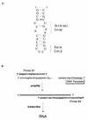
Figure 1
Figure 1
(A) The structure of the simplified 27-nt A-site 16S ribosomal RNA used by Fourmy et al. (22). This structure is employed as the target for the in vitro selection protocol. (B) The nucleotide sequences of the 109mer and 69mer DNA template have embedded within them a 60 random nt and a 20 random nt cassette, respectively. The DNA templates are then amplified through seven cycles of PCR using primers #1 and #2. Primer #2 has a promoter sequence for T7 polymerase reactions (sequence in italic). The dsDNA was then reverse transcribed to the corresponding RNA using the T7 transcription kit from Promega.
Figure 2
Figure 2
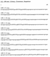
Randomized nucleotide sequences of the selected RNA aptamer constructs from the (A) 109mer and (B) 69mer libraries that exhibit high specificity and affinity towards the A-site 16S rRNA construct. The consensus sequences within each library are highlighted in bold lettering.
Figure 2
Figure 2
Randomized nucleotide sequences of the selected RNA aptamer constructs from the (A) 109mer and (B) 69mer libraries that exhibit high specificity and affinity towards the A-site 16S rRNA construct. The consensus sequences within each library are highlighted in bold lettering.
Figure 3
Figure 3
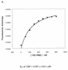
(A) The structure of the fluorescein-labeled 27-nt A-site 16S ribosomal RNA utilized in the fluorescence measurements. (B) Fluorescence anisotropy of fluorescein-labeled A-site 16S rRNA as a function of the 109.2-3 aptamer concentrations. Curve fitting (solid line) using equation 1 gave Kd = 1.236 µM.
Figure 3
Figure 3
(A) The structure of the fluorescein-labeled 27-nt A-site 16S ribosomal RNA utilized in the fluorescence measurements. (B) Fluorescence anisotropy of fluorescein-labeled A-site 16S rRNA as a function of the 109.2-3 aptamer concentrations. Curve fitting (solid line) using equation 1 gave Kd = 1.236 µM.
Figure 4
Figure 4
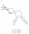
The secondary structure of the 109mer aptamer 109.2-3 as predicted by the Mfold program (30). The circled nucleotides indicate the consensus sequences of the aptamer.
Figure 5
Figure 5
(A) Fluorescence anisotropy of CRP (10 nM) as a function of the simplified 16S rRNA analogue concentrations. Curve fitting (solid line) using equation 1 gave Kd = 0.207 µM. (B) Fluorescence anisotropy of CRP (10 nM) containing the 16S rRNA analogue as a function of neomycin B concentrations. Curve fitting (solid line) using equation 2 gave Kd = 0.248 µM.
Figure 5
Figure 5
(A) Fluorescence anisotropy of CRP (10 nM) as a function of the simplified 16S rRNA analogue concentrations. Curve fitting (solid line) using equation 1 gave Kd = 0.207 µM. (B) Fluorescence anisotropy of CRP (10 nM) containing the 16S rRNA analogue as a function of neomycin B concentrations. Curve fitting (solid line) using equation 2 gave Kd = 0.248 µM.
Figure 6
Figure 6
(A) Fluorescence anisotropy of CRP (10 nM) containing the A-site 16S rRNA construct as a function of the RNA aptamer 109.2-3 concentrations. (B) Fluorescence anisotropy of fluorescein-labeled A-site 16S rRNA construct (10 nM) containing the RNA aptamer 109.2-3 as a function of the neomycin aminoglycoside concentrations.
Figure 6
Figure 6
(A) Fluorescence anisotropy of CRP (10 nM) containing the A-site 16S rRNA construct as a function of the RNA aptamer 109.2-3 concentrations. (B) Fluorescence anisotropy of fluorescein-labeled A-site 16S rRNA construct (10 nM) containing the RNA aptamer 109.2-3 as a function of the neomycin aminoglycoside concentrations.
Figure 7
Figure 7
(A) Sequence of the antisense DNA oligomer to the consensus stem–loop of aptamer 109.2-3. (B) Minimized structure of the A-site 16S rRNA decoding region stem–loop mimic constructs as determined by the Mfold program (30).
Figure 8
Figure 8
(A) Fluorescence anisotropy of fluorescein-labeled A-site 16S rRNA as a function of the antisense DNA oligomer–109.2-3 aptamer complex concentrations. (B) Fluorescence anisotropy of fluorescein-labeled A-site 16S rRNA as a function of the RNA oligomer–109.2-3 aptamer concentrations. Open squares, incubation of aptamer 109.2-3 with 1 equivalent of construct A; open diamonds, incubation of aptamer 109.2-3 with 2 equivalents of construct A; closed circles, incubation of aptamer 109.2-3 with 1 equivalent of construct B.
Figure 8
Figure 8
(A) Fluorescence anisotropy of fluorescein-labeled A-site 16S rRNA as a function of the antisense DNA oligomer–109.2-3 aptamer complex concentrations. (B) Fluorescence anisotropy of fluorescein-labeled A-site 16S rRNA as a function of the RNA oligomer–109.2-3 aptamer concentrations. Open squares, incubation of aptamer 109.2-3 with 1 equivalent of construct A; open diamonds, incubation of aptamer 109.2-3 with 2 equivalents of construct A; closed circles, incubation of aptamer 109.2-3 with 1 equivalent of construct B.
Figure 9
Figure 9
(A) Point mutations were performed on each of the three nucleotides in the consensus sequence confined to the stem–loop region. The structural integrity of the resulting constructs 1, 2 and 3 remained unchanged as predicted by the Mfold program (29). (B) Fluorescence anisotropy of fluorescein-labeled A-site 16S rRNA as a function of the three mutant 109.2-3 aptamer construct concentrations. Open diamonds, construct 1; crossed square boxes, construct 2; closed triangles, construct 3; closed circles, wild type aptamer 109.2-3 construct.
Figure 9
Figure 9
(A) Point mutations were performed on each of the three nucleotides in the consensus sequence confined to the stem–loop region. The structural integrity of the resulting constructs 1, 2 and 3 remained unchanged as predicted by the Mfold program (29). (B) Fluorescence anisotropy of fluorescein-labeled A-site 16S rRNA as a function of the three mutant 109.2-3 aptamer construct concentrations. Open diamonds, construct 1; crossed square boxes, construct 2; closed triangles, construct 3; closed circles, wild type aptamer 109.2-3 construct.

References

    1. Ellington A.D. and Szostak,J.W. (1990) Nature, 346, 818–822. - PubMed
    1. Tuerk C. and Gold,L. (1990) Science, 249, 505–510. - PubMed
    1. Beaudry A. and Joyce,G.F. (1992) Science, 257, 635–641. - PubMed
    1. Gold L., Polisky,B., Uhlenbeck,O. and Yarus,M. (1995) Annu. Rev. Biochem., 64, 763–797. - PubMed
    1. Pei D., Ulrich,H.D. and Schultz,P.G. (1991) Science, 253, 1408–1411. - PubMed

Publication types