. 2000 Aug 1;28(15):2959-68.
doi: 10.1093/nar/28.15.2959.

Molecular characterization at the RNA and gene levels of U3 snoRNA from a unicellular green alga, Chlamydomonas reinhardtii

Affiliations

Molecular characterization at the RNA and gene levels of U3 snoRNA from a unicellular green alga, Chlamydomonas reinhardtii

M Antal et al. Nucleic Acids Res. .

Abstract

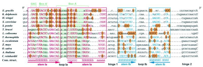
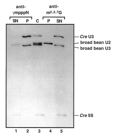
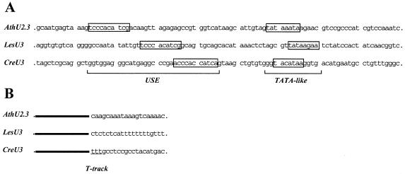
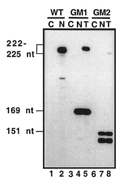
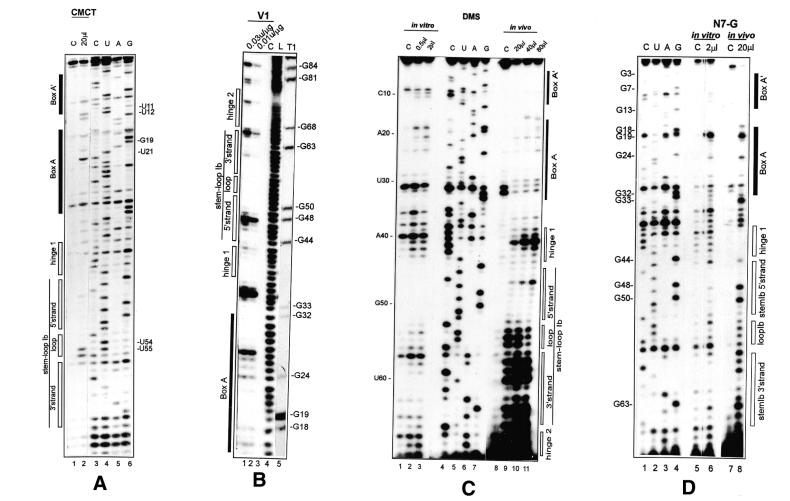
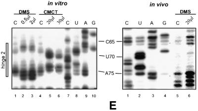
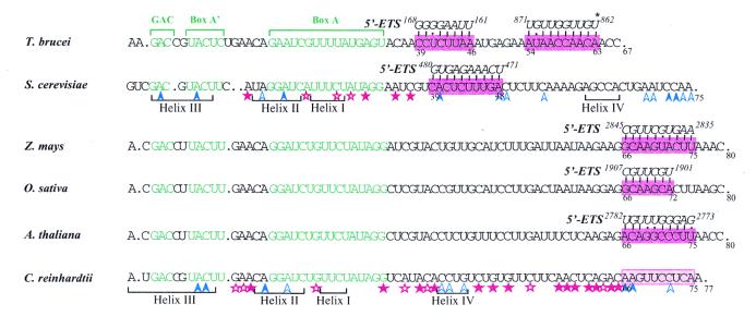
A U3 snoRNA gene isolated from a Chlamydomonas reinhardtii (CRE:) genomic library contains putative pol III-specific transcription signals similar to those of RNA polymerase III-specific small nuclear (sn)RNA genes of higher plants. The 222 nt long CRE: U3 snoRNA was immunoprecipitated by anti-gamma-mpppN antisera, but not by anti-m(2,2,7)G antibodies, supporting the notion that it is a RNA polymerase III transcript. Tagged CRE: U3 snoRNA gene constructs were expressed in CRE: cells. Results of chemical and enzymatic structure probing of CRE: U3 snoRNA in solution and of DMS modification of CRE: U3 snoRNA under in vivo conditions revealed that the two-hairpin structure of the 5'-domain that is found in solution is no longer detected under in vivo conditions. The observed differences can be explained by the formation of several base pair interactions with the 18S and 5'-ETS parts of the pre-rRNA. A model that involves five intermolecular helices is proposed.


Figures

Figure 1
Figure 1
Compensatory base mutations conserved the possibility of forming a two stem–loop structure in yeast, ciliate, unicellular algal and plant U3 snoRNAs. The sequences of the 5′-terminal domain from Euglena gracilis (Eg27297), Kluyveromyces delphensis (Kdz78433), Hansenula wingei (Hwu3sno), Saccharomyces cerevisiae (Scsnr17a), Schizosaccharomyces pombe (Spsnru3), Trypanosoma brucei (Tburb), Leptomonas collosoma (Lctgv3snr), Tetrahymena thermophila (Ttsnru31), Lycopersicon esculentum (Leu3snr), Zea mays (Zmu3snrng), Oryza sativa (Osu3snrn), Arabidopsis thaliana (Atu3csnr) and Chlamydomonas reinhardtii (Crj001179) were first subjected to the program developed by Lück et al. (31). Then alignment was manually refined for a better alignment of the phylogenetically conserved boxes GAC, A′ and A (highlighted in green) and for a better representation of co-variations. The nucleotide sequences involved in the stem of stem–loop Ia are boxed in red. Stems are marked below by inverted arrows. The base paired residues are in capital letters. Red capital letters correspond to residues fitting the consensus sequence established from the alignment. Nucleotide variations from this consensus sequence that were found to preserve base pair interactions are circled in yellow. Dots indicate missing nucleotides as referred to the alignment. The same representation is used for stem–loop Ib, except that blue is used instead of red. The sequences can be accessed via the EMBL-ID at http://srs.ebi.ac.uk:5000/site
Figure 2
Figure 2
Cap structure of Cre U3 snoRNA. U3 snoRNA from Cre and from broad bean, U2 snRNA from broad bean and 5S RNA from Cre were 3-end-labeled, mixed and run on a 10% polyacrylamide gel under semi-denaturing conditions either directly (C, lane 3) or after immunoprecipitation with anti-γmpppN (lanes 1 and 2) or anti-m2,2,7G (lanes 4 and 5) antibodies. RNAs present in the precipitates (P) and in the supernatants (SN) are shown in lanes 2 and 4 and lanes 1 and 5, respectively. Their identities are indicated on the right side of the figure.
Figure 3
Figure 3
Comparative analysis of the Cre U3 gene. The upstream (A) and downstream (B) non-coding regions of the Cre U3 gene that we isolated were aligned with those of the A.thaliana (AthU2.3) (55) and L.esculentum (LesU3) (48) U3 genes. In (A) the upstream (USE) and TATA-like promoter elements identified for the AthU2.3 and LesU3 genes are boxed. A sequence of the Cre U3 gene showing 72.7% identity with the AthU2.3 and LesU3 USE elements is boxed as well as a putative TATA box. In (B) the track of T residues following the Cre U3 coding sequence is underlined.
Figure 4
Figure 4
Expression of Cre U3 snoRNA genes in Cre cells analyzed by RNase A/T1 mapping. Cre cells were transformed with plasmid pCRU3GM1 or pCRU3GM2, coding for a Cre U3 snoRNA (225 nt) with a substitution in loop Ib (positions 53–56) and a Cre U3 snoRNA (223 nt) with a substitution in sequence 70–74, respectively (see Materials and Methods). RNAs extracted from non-transformed cells (N, lanes 2, 4 and 7) and transformed cells (T, lanes 5 and 8), as well as carrier tRNA from E.coli used as a control (C, lanes 1, 3 and 6), were hybridized with uniformly labeled probes prepared by transcription of plasmid pCRU3G (WT, lanes 1 and 2), pCRU3GM1 (GM1, lanes 3–5) or pCRU3GM2 (GM2, lanes 6–8). After RNase A/T1 digestion, the resistant fragments from the RNA probes were analyzed by electrophoresis on a 7% polyacrylamide gel. In lane 2, the wild-type probe (222 nt in length) is protected by the endogenous U3. In lanes 4 and 5, a 165 nt long fragment resulted from partial protection of the mutated probe by endogenous U3 and in lane 5, a full-length probe resulted from protection by the expressed GM1 U3 mutant RNA (225 nt). In lanes 7 and 8, the 150 nt long fragment corresponds to partial protection of the GM2 probe by endogenous U3. In lane 8, the full-length probe was only protected in tiny amounts, indicating a low level of GM2 mutant U3 snoRNA (223 nt) in the transformed cells.
Figure 5
Figure 5
Secondary structure analysis of the Cre U3 snoRNA 5′-domain in solution (A–E, lanes marked in vitro) and in vivo (C–E, lanes marked in vivo). Chemical reagents and enzymes were as described in Materials and Methods. Positions of cleavages and modifications were identified by extension of primer P/Internal Loop (AD) or P/BoxB (E) with reverse transcriptase. The cDNAs were fractionated on a 7% sequencing gel. The amounts of chemical reagents (DMS and CMCT) and enzymes (RNases V1 and T1) used are indicated at the top of the Figure. Control lanes (C) correspond to primer extensions made on RNA incubated in the absence of chemical reagent or enzyme. Lanes C, U, A and G correspond to the sequencing ladder obtained with the same primer. Nucleotide positions in Cre U3 and secondary structural elements are marked on the left and right sides of the pictures.
Figure 5
Figure 5
Secondary structure analysis of the Cre U3 snoRNA 5′-domain in solution (A–E, lanes marked in vitro) and in vivo (C–E, lanes marked in vivo). Chemical reagents and enzymes were as described in Materials and Methods. Positions of cleavages and modifications were identified by extension of primer P/Internal Loop (AD) or P/BoxB (E) with reverse transcriptase. The cDNAs were fractionated on a 7% sequencing gel. The amounts of chemical reagents (DMS and CMCT) and enzymes (RNases V1 and T1) used are indicated at the top of the Figure. Control lanes (C) correspond to primer extensions made on RNA incubated in the absence of chemical reagent or enzyme. Lanes C, U, A and G correspond to the sequencing ladder obtained with the same primer. Nucleotide positions in Cre U3 and secondary structural elements are marked on the left and right sides of the pictures.
Figure 6
Figure 6
Schematic representation of the results of chemical and enzymatic probing on the secondary structure proposed for the Cre U3 snoRNA 5′-domain. (A) In vitro probing. The phylogenically conserved boxes A′ and A are framed in black. Nucleotides modified by DMS or CMCT at Watson–Crick positions are circled. N7-G methylations are shown by black dots; the number of dots reflects the intensity of the modification. Positions of RNase V1 cleavages are indicated by arrows linked to squares. Intensity of the colors (green for chemicals, orange for RNase V1) indicates the intensity of the modification or yield of cleavage. Crosses (×) indicate pauses of the reverse transcriptase. (B) In vivo probing. The code for DMS modification is as in (A). In addition, extremely strong modifications are shown in black. Decreased sensitivity to DMS in vivo as compared to in vitro is indicated by a blue arrow: moderate protection by an open arrow, strong protection by a full arrow. Increased sensitivity is indicated by a purple star: a moderate increase by an open star, a strong increase by a full star. Unusual modifications of U residues are indicated by purple triangles (see text for explanation). The hinge 2 sequence proposed to be complementary to the 5′-ETS rRNA is framed in pink. (C and D) Two alternative models of the Cre U3 snoRNA–pre-rRNA interaction. Bimolecular helices I–IV between the Cre U3 snoRNA (sequence in red) and the Cre 18S rRNA (sequence in blue) according to Hughes (15) and Méreau et al. (14) are shown. The phylogenetically conserved boxes A and A′ are boxed in black. Variation of reactivity of nucleotides to DMS in vivo, as compared to in vitro, is indicated by the same code as in (B).
Figure 7
Figure 7
Alignment of the 5′-portion of U3 snoRNAs from T.brucei, S.cerevisiae, Z.mays, O.sativa, A.thaliana and C.reinhardtii showing sequence complementarities with the 5′-ETS regions of pre-rRNAs. Database accession numbers of the U3 snoRNA sequences are indicated in the legend to Figure 1. The phylogenetically conserved boxes GAC, A′ and A are shown in green. The complementary 5′-ETS sequences are in italic and positions of their extremities as referred to the pre-rRNA transcription start site are indicated. The two complementary 5′-ETS sequences given for T.brucei were proposed by Hartshorne and Tokoyufu (68), U862 from the 5′-ETS shown to be cross-linked to U3 snoRNA is marked by an asterisk. The S.cerevisiae 5′-ETS complementarity corresponds to the well-documented helix V (12). Positions of helices I–IV, formed with the 18S part of the pre-rRNA, are shown, as well as increased (red stars) or decreased (blue arrows) sensitivity of A and C residues to DMS in vivo (14). The U3–5′-ETS complementarities proposed for Z.mays, O.sativa and A.thaliana were obtained by a search performed with pattern(n) software (Infobiogen). For C.reinhardtii, the variations of DMS reactivities observed in the present paper are represented by the same code as for S.cerevisiae. Sequences that may form bimolecular helices I–IV with the 18S part of pre-rRNA are indicated, as well as the potential site of interaction with the 5′-ETS region (highlighted in pink).

Similar articles

Cited by

References

    1. Eichler D.C. and Craig,N. (1994) Prog. Nucleic Acid Res. Mol. Biol., 49, 197–239. - PubMed
    1. Maxwell E.S. and Fournier,M.J. (1995) Annu. Rev. Biochem., 64, 897–934. - PubMed
    1. Raué H.A. and Planta,R.J. (1995) Gene Expr., 5, 71–77. - PMC - PubMed
    1. Bachellerie J.P. and Cavaillé,J. (1998) In Grosjean,H. and Benne,R. (eds), The Modification and Editing of RNA. ASM Press, Washington, DC, pp. 255–272.
    1. Ofengand J. and Fournier,M.J. (1998) In Grosjean,H. and Benne,R. (eds), The Modification and Editing of RNA. ASM Press, Washington, DC, pp. 229–253.

Publication types

MeSH terms

Associated data