. 2000 Aug 1;28(15):2986-92.
doi: 10.1093/nar/28.15.2986.

Compaction of DNA in an anionic micelle environment followed by assembly into phosphatidylcholine liposomes

Affiliations

Compaction of DNA in an anionic micelle environment followed by assembly into phosphatidylcholine liposomes

E A Murphy et al. Nucleic Acids Res. .

Abstract

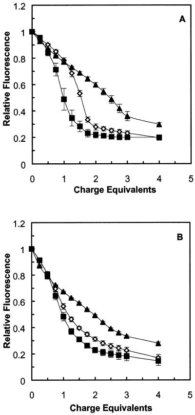
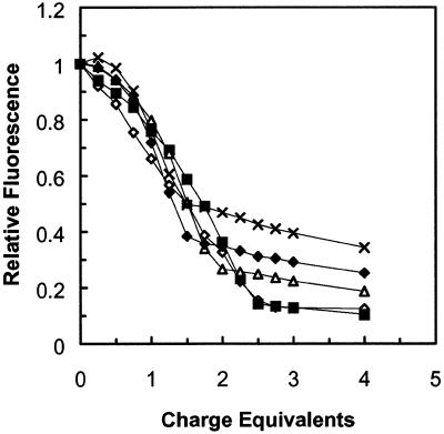
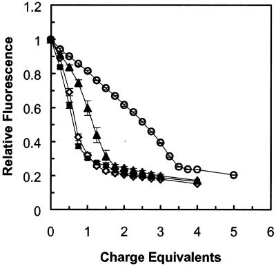
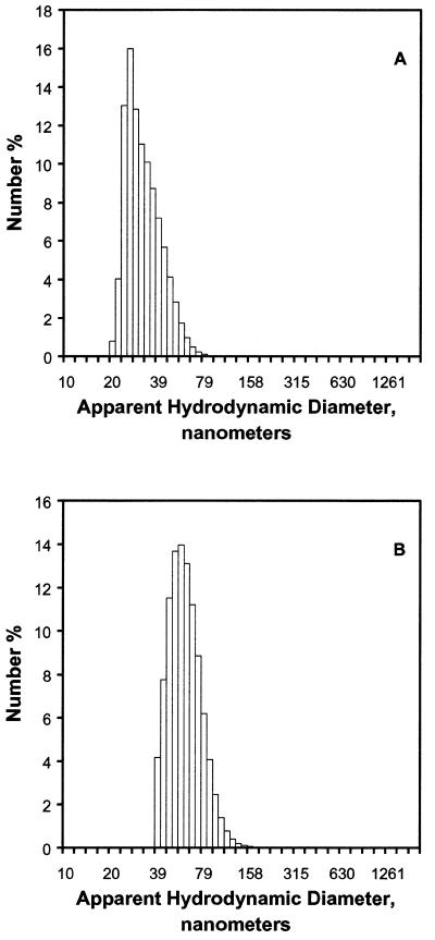
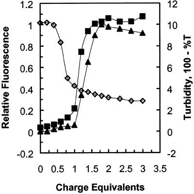
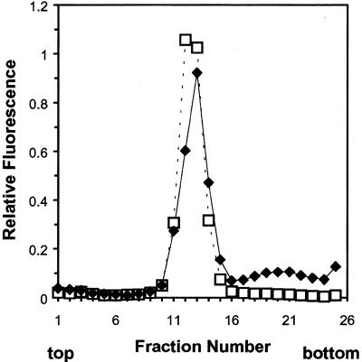
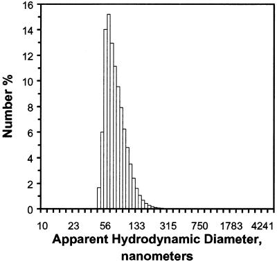
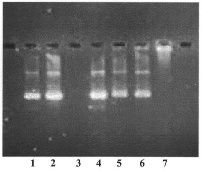
A difficult problem concerning the interaction of DNA with amphiphiles of opposite charge above their critical micelle concentration is the propensity for aggregation of the condensed DNA complexes. In this study, this problem was addressed by attenuating amphiphile charge density within a cholate micelle environment. The amphiphile consisted of a cationic peptide, acetyl-CWKKKPKK-amide, conjugated to dilaurylphos-phatidylethanolamine. In the presence of cholate, multiple equivalents of cationic charge were required to bring about the completion of DNA condensation. At the end point of condensation, stable, soluble DNA-micelle complexes were formed, which by dynamic light scattering exhibited apparent hydro-dynamic diameters between 30 and 60 nm. Aggregation, as measured by static light scattering at 90 degrees and by turbidity, was not observed until further additions of peptide-lipid conjugate were made beyond the end point of DNA condensation. Liposome complexes containing the non-aggregated, compacted DNA were formed by adding dioleoylphosphatidylcholine followed by removing the cholate by dialysis. The resulting complexes were distributed within a narrow density range, the DNA was quantitatively assembled into the liposomes, and liposomes without DNA were not detected. Small particles were formed with a mean hydrodynamic diameter of 77 nm. The liposomal DNA showed complete retention of its supercoiled form and no detectable sensitivity to DNase (25 U/10 microg DNA, 1.5 h, 37 degrees C). The use of an anionic, dialyzable amphiphile to attenuate charge inter-actions between DNA and cationic amphiphiles is a useful technology for the quantitative assembly of compacted DNA into conventional liposomes, with complete protection against nuclease activity.


Figures

Figure 1
Figure 1
DNA condensation by free peptide and peptide–lipid conjugates in anionic detergent. Experimental solutions consisted of 10 µg DNA in 3 ml of 20 mM cholate, 1 mM phosphate buffer, pH 8.0. Solutions of peptide or peptide–lipid conjugate in dimethylformamide were added in 5 µl aliquots (1.5 nmol peptide, 0.25 charge equivalents relative to the DNA). The fluorescence values were normalized to a range of 0–1 as described in Materials and Methods. (A) Ethidium bromide fluorescence, dye:base pair ratio 1:25. Closed triangles, free peptide; open diamonds, C12–PE–peptide; closed squares, C18–PE–peptide. (B) Hoechst dye fluorescence, dye:base pair ratio 1:25. Symbols as in (A). Values are means ± SEM for three separate experiments (for many data points, error bars are eclipsed by the size of the symbol).
Figure 2
Figure 2
DNA condensation by C12–PE peptide–lipid conjugate at different DNA concentrations. Experimental solutions consisted of 20 mM cholate, 1 mM phosphate buffer, pH 8.0, with 2–30 µg DNA in a total volume of 3 ml. The ethidium bromide to DNA base pair ratio was maintained at 1:25 mol/mol regardless of DNA concentration. C12–PE peptide–lipid conjugate in dimethylformamide was added in 5 µl aliquots, 0.25 charge equivalents per aliquot. Observed fluorescence was normalized to a range of 0–1 as described in Materials and Methods. Open diamonds, 2 µg DNA; closed squares, 5 µg DNA; open triangles, 10 µg DNA; closed diamonds, 20 µg DNA; crosses, 30 µg DNA.
Figure 3
Figure 3
DNA condensation by C12–PE peptide–lipid conjugate at different concentrations of sodium cholate. Experimental solutions consisted of 1 mM phosphate buffer, pH 8.0, 10 µg DNA, 3 ml total volume, and cholate concentrations of 10–30 mM. C12–PE peptide–lipid conjugate in dimethylformamide was added in 5 µl aliquots, 0.25 charge equivalents per aliquot. Observed fluorescence was normalized to a range of 0–1 as described in Materials and Methods. Values are means ± SEM for three separate experiments (for most data points, error bars are eclipsed by the size of the symbol). Open diamonds, 10 mM cholate; closed squares, 15 mM cholate; closed triangles, 20 mM cholate; open circles, 30 mM cholate.
Figure 4
Figure 4
Dynamic light scattering of condensed DNA in 20 mM cholate at two different DNA concentrations. (A) Five micrograms plasmid DNA condensed with 2.5 charge equivalents of C12–PE peptide–lipid conjugate, 3 ml total volume. (B) Ten micrograms DNA condensed with 2.0 charge equivalents of peptide–lipid conjugate, 3 ml total volume.
Figure 5
Figure 5
Determination of the charge equivalence difference between the end point of DNA condensation and the onset of aggregation and turbidity in anionic detergent. Experimental solutions consisted of 1 mM phosphate buffer, pH 8.0, 10 µg DNA, 3 ml total volume. Cholate concentration was 20 (A) or 30 mM (B). DNA condensation (closed diamonds) was measured by ethidium bromide displacement and observed fluorescence was normalized to a range of 0–1 as described in Materials and Methods. Aggregation was measured by static light scattering at 90° (closed squares) or by turbidity (open triangles), as described in Materials and Methods. C12–PE peptide–lipid conjugate in dimethylformamide was added in 5 µl aliquots, 0.25 charge equivalents per aliquot, in the experiment in (A), and 5 µl aliquots, 0.33 charge equivalents per aliquot, for the experiment in (B).
Figure 6
Figure 6
Determination of the charge equivalence difference between the end point of DNA condensation and the onset of aggregation and turbidity in neutral detergent. Experimental solutions consisted of 1 mM phosphate buffer, pH 8.0, 10 µg DNA, 3 ml total volume. Octyl glucoside concentration was 30 mM. DNA condensation (open diamonds) was measured by ethidium bromide displacement and observed fluorescence was normalized to a range of 0–1 as described in Materials and Methods. Aggregation was measured by static light scattering at 90° (closed squares) or by turbidity (closed triangles), as described in Materials and Methods. C12–PE peptide–lipid conjugate in dimethylformamide was added in 5 µl aliquots, 0.20 charge equivalents per aliquot.
Figure 7
Figure 7
Density gradient analysis of DNA–phosphatidylcholine liposomes. Liposomes containing 0.1% of the fluorescent lipid 1-(18:1)-2-(C16-BODIPY)-phosphatidylcholine were concentrated to ∼20 µg DNA/ml and a 0.5 ml aliquot placed on a 4 ml 0–20% linear sucrose gradient. Fractions of 150 µl were collected then diluted to 1 ml with 1 mM phosphate buffer, pH 8.0. Lipid content (open squares) was measured by fluorescence (λex 490 nm, λem 520 nm). DNA (closed diamonds) was measured by the addition of Hoechst dye (λex 350 nm, λem 460 nm).
Figure 8
Figure 8
Dynamic light scattering analysis of DNA–phosphatidylcholine liposomes. Liposome composition, method of assembly and instrumental details of the analysis are described in Materials and Methods.
Figure 9
Figure 9
DNase resistance of DNA–phosphatidylcholine liposomes. Liposomes containing 10 µg DNA were treated with 25 U DNase for 1.5 h at 37°C as described in Materials and Methods. Lipid and peptide–lipid conjugate were extracted into chloroform as described and the DNA (which remained in the aqueous phase) was analyzed on a 1% agarose gel containing ethidium bromide (∼1 µg/lane). Lane 1, free DNA; lane 2, free DNA taken through the extraction procedure (no DNase); lane 3, free DNA completely digested by DNase; lane 4, a ‘stop’ control, where free DNA was added after an incubation mixture containing DNase had been extracted with chloroform; lane 5, DNA–phosphatidylcholine liposomes incubated without DNase and carried through the extraction procedure; lane 6, DNA–phosphatidylcholine liposomes incubated with DNase and carried through the extraction procedure; lane 7, DNA–phosphatidylcholine liposomes not treated with DNase and not extracted.

References

    1. Fraley R. and Papahadjopoulos,D. (1982) Curr. Top. Microbiol. Immunol., 96, 171–191. - PubMed
    1. Lurquin P.F. (1993) In Gregoriadis,G. (ed.), Liposome Technology, Vol. II, 2nd Edn. CRC Press, Boca Raton, FL, pp. 129–139.
    1. Felgner P.L., Gadek,T.R., Holm,M., Roman,R., Chan,H.W., Wenz,M., Northrop,J.P., Ringold,G.M. and Danielsen,M. (1987) Proc. Natl Acad. Sci. USA, 84, 7413–7417. - PMC - PubMed
    1. Felgner P.L., Barenholz,Y., Behr,J.P., Cheng,S.H., Cullis,P., Huang,L., Jessee,J.A., Seymour,L., Szoka,F. and Thierry,A.R. (1997) Hum. Gene Ther., 8, 511–512. - PubMed
    1. Lasic D.D. (1997) Liposomes in Gene Delivery. CRC Press, Boca Raton, FL.