. 2000 Jul 24;150(2):349-60.
doi: 10.1083/jcb.150.2.349.

HIV-1 rev depolymerizes microtubules to form stable bilayered rings

Affiliations

HIV-1 rev depolymerizes microtubules to form stable bilayered rings

N R Watts et al. J Cell Biol. .

Abstract

We describe a novel interaction between HIV-1 Rev and microtubules (MTs) that results in the formation of bilayered rings that are 44-49 nm in external diameter, 3.4-4.2 MD (megadaltons) in mass, and have 28-, 30-, or 32-fold symmetry. Ring formation is not sensitive to taxol, colchicine, or microtubule-associated proteins, but requires Mg(2+) and is inhibited by maytansine. The interaction involves the NH(2)-terminal domain of Rev and the face of tubulin exposed on the exterior of the MTs. The NH(2)-terminal half of Rev has unexpected sequence similarity to the tubulin-binding portion of the catalytic/motor domains of the microtubule-destabilizing Kin I kinesins. We propose a model wherein binding of Rev dimers to MTs at their ends causes segments of two neighboring protofilaments to peel off and close into rings, circumferentially containing 14, 15, or 16 tubulin heterodimers, with Rev bound on the inside. Rev has a strong inhibitory effect on aster formation in Xenopus egg extracts, demonstrating that it can interact with tubulin in the presence of normal levels of cellular constituents. These results suggest that Rev may interact with MTs to induce their destabilization, a proposition consistent with the previously described disruption of MTs after HIV-1 infection.


Figures

Figure 1
Figure 1
Rev–tubulin rings and related complexes. Shown are Rev filaments (A), MTs (B), colchicine depolymerized tubulin (C), RTTs (D), RTTs forming at the ends of MTs (E), Rev 1–59–tubulin rings (F), and Dolastatin-10–tubulin rings (G). RTT double-bars are visible in D (arrows). The inset in E shows MTs depolymerizing at both ends. Bars: (A–G) 100 nm; (inset) 200 nm.
Figure 2
Figure 2
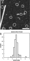
Mass distribution of RTTs determined by STEM. Shown are a field of RTTs and a TMV particle (A), and the mass histogram (B). Bar, 100 nm.
Figure 3
Figure 3
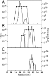
Detection of rotational symmetries in RTTs as a function of radius. Two measures of significance, Student's t test and the ratio product (Kocsis et al. 1995), were applied to two subsets of images, comprising 167 smaller rings and 256 larger rings, respectively. For polar-sampled images, the t test assesses at each radius and for each potential order of symmetry, whether that symmetry is significantly represented in the data set as a whole. The only significant symmetries detected were 28-fold (A) in the smaller set, and 30-fold (B), and 32-fold (C) in the larger set. For instance, in A, 28-fold symmetry is detected at the P < 10−6 level at radii of 18–22 nm. N-fold symmetry at radius R corresponds to an azimuthal spacing of 2πR/N in the specimen. For a given candidate symmetry at a given radius, the ratio product is the product of the corresponding Fourier amplitudes from all the images, normalized against the average amplitude from the background images. This product converges rapidly to zero except when a certain symmetry is significantly present. The relative proportions of particles with different symmetries may be estimated from the product ratios when both saturate the t test–calculated limit of P < 10−6. In this case, it may be estimated that 30-fold rings are three- to fourfold more abundant in the large subset than are 32-fold rings.
Figure 4
Figure 4
Image analysis and a molecular model of RTTs. Shown are the cross-correlation averages of 28-, 30-, and 32-fold symmetric particles viewed axially (A–C), the corresponding power spectra (D–F), and the final symmetrized images (G–I). Also shown are selected side-views of rings (J), the correlation average (K), and a symmetrized image (L). A molecular model of the 30-fold RTT is shown in M and N. In the model, Rev monomers are located on the inside and tubulin monomers on the outside (M). The side-view (N) shows RTTs as composed of bi-layered rings. The α- and β-tubulin monomers are differentiated as black and white. Bar, 20 nm.
Figure 5
Figure 5
Domain structure and amino acid sequence similarity of HIV-1 Rev to kinesin XKCM1. A shows the locations of Helix 1, the polyproline sequence, Helix 2 (the RNA-binding region with flanking multimerization domains M1 and M2), and the activation domain of Rev. Key residues and positions are numbered above. B shows the XKCM1 motor/catalytic domain sequence from residues 500–550, and aligned Rev sequences below. In Rev, residues are identical (red), very similar (green), or similar (blue) to XKCM1. These residues are bold in XKCM1. Specific residues that are highly conserved in both Rev and the kinesins and that have identified structural and/or functional roles are marked by asterisks. Rev isolates are identified on the right. Rev isolate P04616 (carats) was used in this study.
Figure 6
Figure 6
Inhibition of MT polymerization by Rev in Xenopus egg extracts. Shown is aster formation in the absence (A–C) and presence (D–F) of Rev. Sperm chromatin fluoresces blue (4′,6-diamidino-2-phenylindole dihydrochloride), and Rhodamine-tubulin fluoresces red. Bar, 10 μm.

Similar articles

Cited by

References

    1. Auer M., Gremlich H.-U., Seifert J.-M., Daly T.J., Parslow T.G., Casari G., Gstach H. Helix-loop-helix motif in HIV-1 Rev. Biochemistry. 1994;33:2988–2996. - PubMed
    1. Bai R., Taylor G.F., Schmidt J.M., Williams M.D., Kepler J.A., Pettit G.R., Hamel E. Interaction of dolastatin 10 with tubulininduction of aggregation and binding and dissociation reactions. Mol. Pharmacol. 1995;47:965–976. - PubMed
    1. Bai R., Schwartz R.E., Kepler J.A., Pettit G.R., Hamel E. Characterization of the interaction of cryptophycin 1 with tubulinbinding in the Vinca domain, competitive inhibition of dolastatin 10 binding, and an unusual aggregation reaction. Cancer Res. 1996;56:4398–4406. - PubMed
    1. Bai R., Durso N.A., Sackett D.L., Hamel E. Interactions of the sponge-derived antimitotic tripeptide hemiasterlin with tubulincomparison with dolastatin 10 and cryptophycin 1. Biochemistry. 1999;38:14302–14310. - PubMed
    1. Battiste J.L., Mao H.Y., Rao N.S., Tan R.Y., Muhandiram D.R., Kay L.E., Frankel A.D., Williamson J.R. Alpha helix-RNA major groove recognition in an HIV-1 Rev peptide RRE RNA complex. Science. 1996;273:1547–1551. - PubMed

Publication types

MeSH terms