. 2000 Aug 1;20(15):5696-702.
doi: 10.1523/JNEUROSCI.20-15-05696.2000.

Clinical mutations in the L1 neural cell adhesion molecule affect cell-surface expression

Affiliations

Clinical mutations in the L1 neural cell adhesion molecule affect cell-surface expression

H D Moulding et al. J Neurosci. .

Abstract

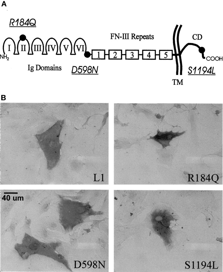
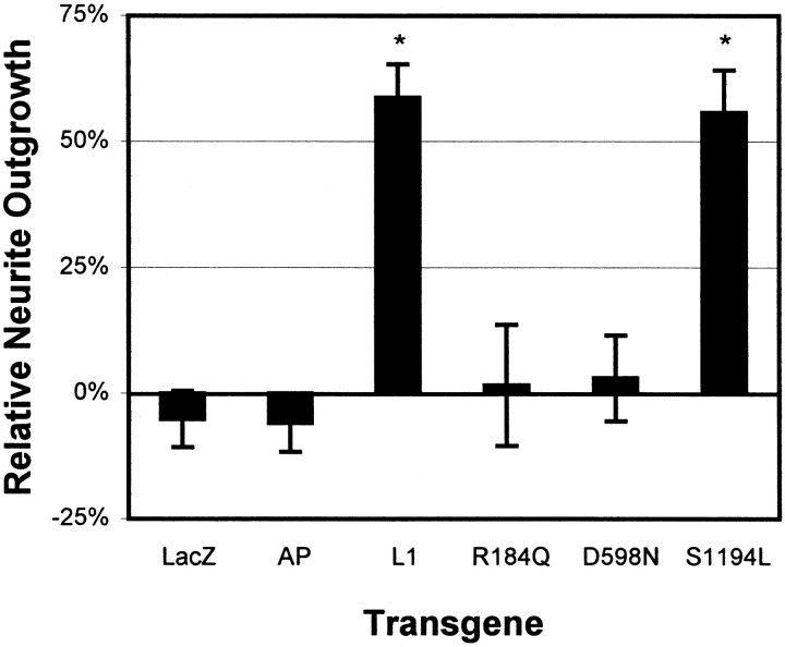
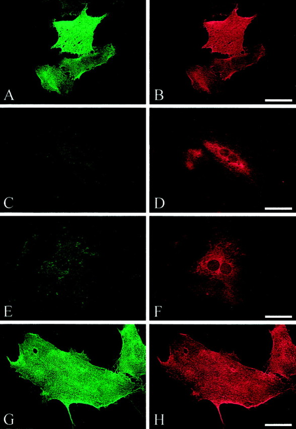
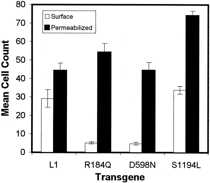
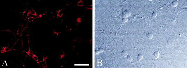
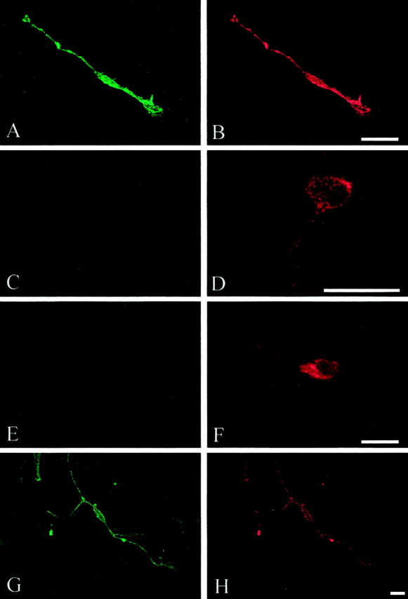
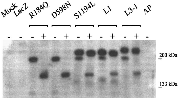
Mutations in the L1 neural cell adhesion molecule, a transmembrane glycoprotein, cause a spectrum of congenital neurological syndromes, ranging from hydrocephalus to mental retardation. Many of these mutations are single amino acid changes that are distributed throughout the various domains of the protein. Defective herpes simplex virus vectors were used to express L1 protein with the clinical missense mutations R184Q and D598N in the Ig2 and Ig6 extracellular domains, respectively, and S1194L in the cytoplasmic domain. All three mutant proteins were expressed at similar levels in infected cells. Neurite outgrowth of cerebellar granule cells was stimulated on astrocytes expressing wild-type or S1194L L1, whereas those expressing R184Q and D598N L1 failed to increase neurite length. Live cell immunofluorescent staining of L1 demonstrated that most defective vector-infected cells did not express R184Q or D598N L1 on their cell surface. This greatly diminished cell-surface expression occurred in astrocytes, neurons, and non-neural cells. In contrast to wild-type or S1194L L1, the R184Q and D598N L1 proteins had altered apparent molecular weights and remained completely endoglycosidase H (endoH)-sensitive, suggesting incomplete post-translational processing. We propose that some missense mutations in human L1 impede correct protein trafficking, with functional consequences independent of protein activity. This provides a rationale for how expressed, full-length proteins with single amino acid changes could cause clinical phenotypes similar in severity to knock-out mutants.


Figures

Fig. 1.
Fig. 1.
A, Schematic of the L1 cell adhesion molecule showing domain structure: Ig-like domains,I–VI; fibronectin-type III repeats 1–5,FN-III; transmembrane region, TM; and cytoplasmic domain, CD. The location of the mutations (underlined and italicized) are indicated (●). B, Immunohistochemical staining of L1 protein in fixed-permeabilized astrocytes after infection with dvL1.
Fig. 2.
Fig. 2.
Wild-type and S1194L mutant L1 stimulate neurite outgrowth. Relative change in mean neurite lengths of CGCs plated on astrocytes infected with dvL1 (wild-type or indicated mutant), dvHCL (LacZ), or dvHC-AP (AP). Data pooled from four separate experiments after normalization to neurite lengths on mock-infected astrocytes. *p < 0.05 by Tukey's studentized range tests conducted after one-way ANOVA (p = 0.0001).
Fig. 3.
Fig. 3.
Surface (A, C,E, G) and permeabilized (B, D, F,H) staining of L1. Double immunofluorescence was performed on rat cerebellar astrocytes infected with dvL1 (16.5 hr after infection): wild type, A, B; R184Q,C, D; D598N, E,F; and S1194L, G, H. In most cells expressing R184Q and D598N mutant L1, L1 protein failed to reach the cell surface. Scale bar, 50 μm.
Fig. 4.
Fig. 4.
Quantification of surface versus permeabilized L1 double labeling on dvL1-infected astrocytes (16.5 hr after infection). Mean ± SE number of positive cells for surface (open bars) or permeabilized (filled bars) staining per row using 20× objective.
Fig. 5.
Fig. 5.
Endogenous L1 expression in rat CGC neurons. L1 was detected by immunofluorescence (A) using a species nonspecific anti-L1 antibody in fixed-permeabilized CGC neurons (B, differential-interface contrast microscopy). Scale bar, 50 μm.
Fig. 6.
Fig. 6.
Surface (A, C,E, G) and permeabilized (B, D, F,H) staining of exogenous L1. Double immunofluorescence was performed on CGC neurons infected with dvL1 (16.5 hr after infection): wild type, A,B; R184Q, C, D; D598N,E, F; and S1194L, G,H. In most cells expressing R184Q and D598N mutant L1, L1 protein failed to reach the cell surface but was detected within the neuronal cytosol, including processes. Scale bar, 20 μm.
Fig. 7.
Fig. 7.
Western blot of lysates from vector-infected astrocytes. Cell lysates were treated with (+) or without (−) endoH to remove unmodified N-linked glycosylations. Five (R184Q or D598N) or 12 (all others, so lower MW band at 200 kDa would be visible) μg of protein were loaded per lane. The upper MW band, the fully glycosylated form of L1 (∼210 kDa), is resistant to endoH (compare − with + in L1, S1194L, and L3–1), whereas the lower MW band of L1 is sensitive.

References

    1. Aridor M, Bannykh SI, Rowe T, Balch WE. Cargo can modulate COPII vesicle formation from the endoplasmic reticulum. J Biol Chem. 1999;274:4389–4399. - PubMed
    1. Asou H, Miura M, Kobayashi M, Uyemura K. The cell adhesion molecule L1 has a specific role in neural cell migration. NeuroReport. 1992;3:481–484. - PubMed
    1. Bateman A, Jouet M, MacFarlane J, Du JS, Kenwrick S, Chothia C. Outline structure of the human L1 cell adhesion molecule and the sites where mutations cause neurological disorders. EMBO J. 1996;15:6050–6059. - PMC - PubMed
    1. Berger J, Garattini E, Hua JC, Udenfriend S. Cloning and sequencing of human intestinal alkaline phosphatase cDNA. Proc Natl Acad Sci USA. 1987;84:695–698. - PMC - PubMed
    1. Brummendorf T, Rathjen FG. Structure/function relationships of axon-associated adhesion receptors of the immunoglobulin superfamily. Curr Opin Neurobiol. 1996;6:584–593. - PubMed

Publication types

MeSH terms

Substances

LinkOut - more resources