. 2000 Aug 1;20(15):5709-14.
doi: 10.1523/JNEUROSCI.20-15-05709.2000.

Ibuprofen suppresses plaque pathology and inflammation in a mouse model for Alzheimer's disease

Affiliations

Ibuprofen suppresses plaque pathology and inflammation in a mouse model for Alzheimer's disease

G P Lim et al. J Neurosci. .

Abstract

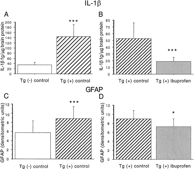
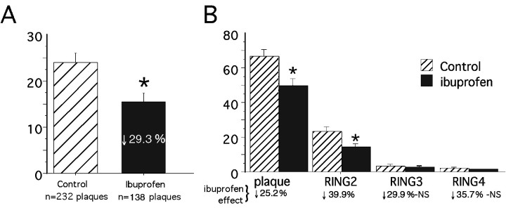
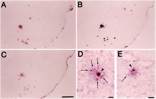
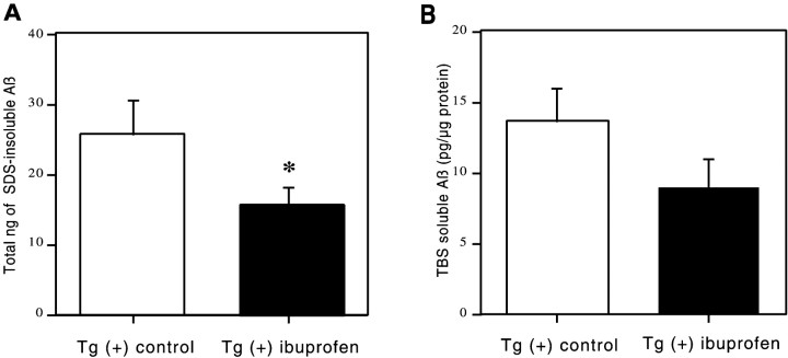
The brain in Alzheimer's disease (AD) shows a chronic inflammatory response characterized by activated glial cells and increased expression of cytokines and complement factors surrounding amyloid deposits. Several epidemiological studies have demonstrated a reduced risk for AD in patients using nonsteroidal anti-inflammatory drugs (NSAIDs), prompting further inquiries about how NSAIDs might influence the development of AD pathology and inflammation in the CNS. We tested the impact of chronic orally administered ibuprofen, the most commonly used NSAID, in a transgenic model of AD displaying widespread microglial activation, age-related amyloid deposits, and dystrophic neurites. These mice were created by overexpressing a variant of the amyloid precursor protein found in familial AD. Transgene-positive (Tg+) and negative (Tg-) mice began receiving chow containing 375 ppm ibuprofen at 10 months of age, when amyloid plaques first appear, and were fed continuously for 6 months. This treatment produced significant reductions in final interleukin-1beta and glial fibrillary acidic protein levels, as well as a significant diminution in the ultimate number and total area of beta-amyloid deposits. Reductions in amyloid deposition were supported by ELISA measurements showing significantly decreased SDS-insoluble Abeta. Ibuprofen also decreased the numbers of ubiquitin-labeled dystrophic neurites and the percentage area per plaque of anti-phosphotyrosine-labeled microglia. Thus, the anti-inflammatory drug ibuprofen, which has been associated with reduced AD risk in human epidemiological studies, can significantly delay some forms of AD pathology, including amyloid deposition, when administered early in the disease course of a transgenic mouse model of AD.


Figures

Fig. 1.
Fig. 1.
Effect of ibuprofen on IL-1β levels and GFAP levels in Tg2576 brains. A, B, IL-1β measurements. ANOVA analyses were performed on measurements in Tg− mice fed control diet (n = 5), Tg+ mice fed control diet (n = 8), and Tg+ mice fed ibuprofen (n = 9). A, Measurement of IL-1β levels in hippocampus and residual cortex in 16-month-old Tg+ and Tg− mice. IL-1β protein levels were measured in TBS-extracted supernatant fractions from Tg− mice fed control diet (open bar) and Tg+ mice fed control diet (hatched bar). Levels were significantly increased in both regions in Tg+ compared to Tg− animals. B, Effect of ibuprofen on IL-1β levels in Tg+ mice. IL-1β was decreased by 64.7% across all regions in ibuprofen-treated animals. Equality of variance was established with a logarithmic transformation. C, D, GFAP levels. C, Effect of transgene on GFAP levels. Semiquantitative measurements of GFAP were performed on immunoblots of Tg− and Tg+ animals fed control diet. A highly significant 51.7% elevation in Tg+ animals was found. D, Effect of ibuprofen on GFAP levels in Tg+ mice. Treatment with ibuprofen significantly decreased GFAP levels 20% across all regions in Tg+ animals. *p < 0.05. ***p < 0.001. Error bars indicate SE.
Fig. 2.
Fig. 2.
Image analysis of phosphotyrosine-labeled microglia. Histograms illustrate the percentage of anti-PT-stained microglial area plus or minus 95% confidence interval in and around Aβ-stained plaque deposits in Tg+ mice on either a control diet (n = 6 mice) or ibuprofen diet (n = 5 mice) in the midhippocampal region (bregma, −1.70 mm). An average of 37 ± 6.0 and 27.35 ± 7.6 plaques were counted per mouse in the control and ibuprofen diets, respectively. A illustrates that ibuprofen induced a 29.3% reduction in the percentage of PT-stained area within four plaque radii from Aβ-stained plaque center, compared to the control diet (p < 0.0001). Logarithmic transformation was needed to establish homogeneity of variance.B, Two-way ANOVA (treatment × ring) showed significant treatment and ring effects as well as a significant treatment–ring interaction (*p < 0.0001). This histogram shows the greater reduction in PT-stained microglia in radii outside the plaque compared to within the plaque (each ring corresponds to one plaque radii). NS, Not statistically significant.
Fig. 3.
Fig. 3.
Representative examples of immunostaining of piriform cortex/amygdala regions from Tg+ mice fed control diet.A and B show staining with anti-Aβ34–40 (A) and DAE (B). Magnification for both is 25×. These antibodies are rabbit polyclonal antisera made to synthetic peptides representing Aβ 34–40 (Mak et al. 1994) and Aβ 1–13, respectively. C shows staining with a rabbit polyclonal antibody made to ubiquitin conjugates (Dako; magnification 25×). Tg+ mice fed control diet (D, magnification 100×) and Tg+ mice fed ibuprofen diet (E, magnification 132×) show double labeling with a monoclonal anti-phosphotyrosine (brown) followed by a light development of DAE (blue). Arrowheads depict microglia (within Aβ-labeled plaque), whereas arrows show microglia surrounding the plaque.
Fig. 4.
Fig. 4.
ELISA measurement of water-soluble and SDS-insoluble Aβ. These histograms illustrate Aβ levels plus or minus 95% confidence interval of formic acid-extracted (SDS-insoluble) Aβ (nanograms per total pellet) or soluble Aβ (picograms per microgram of protein) as measured by sandwich ELISA for dissected brain regions of the Tg2576 brain. A two-way ANOVA (treatment × region) showed significant treatment effects in insoluble Aβ levels (*p < 0.05) and regional effects (p < 0.0001) with no treatment–region interaction. Decreases in soluble Aβ levels were consistent in all regions but did not quite reach statistical significance (p = 0.06).

References

    1. Ard MD, Cole GM, Wei J, Mehrle AP, Fratkin JD. Scavenging of Alzheimer's amyloid beta-protein by microglia in culture. J Neurosci Res. 1996;43:190–202. - PubMed
    1. Benzing WC, Wujek JR, Ward EK, Shaffer D, Ashe KH, Younkin SG, Brunden KR. Evidence for glial-mediated inflammation in aged APPSw transgenic mice. Neurobiol Aging. 2000;20:581–589. - PubMed
    1. Breitner JCS, Welsh KA, Helms MJ, Gaskell PC, Gau BA, Roses AD, Pericak-Vance MA, Saunders AM. Delayed onset of Alzheimer's disease with nonsteroidal anti-inflammatory and histamine H2 blocking drugs. Neurobiol Aging. 1995;16:523–530. - PubMed
    1. Combs CK, Johnson DE, Karlo JC, Cannady SB, Landreth GE. Inflammatory mechanisms in Alzheimer's disease: inhibition of β-amyloid-stimulated proinflammatory responses and neurotoxicity by PPARγ agonists. J Neurosci. 2000;20:558–567. - PMC - PubMed
    1. Dickson DW, Lee SC, Mattiace LA, Yen S-HC, Brosnan C. Microglia and cytokines in neurological disease, with special reference to AIDS and Alzheimer's disease. Glia. 1993;7:75–83. - PubMed

Publication types

MeSH terms