. 2000 Aug 1;20(15):5724-32.
doi: 10.1523/JNEUROSCI.20-15-05724.2000.

The SNARE Vti1a-beta is localized to small synaptic vesicles and participates in a novel SNARE complex

Affiliations

The SNARE Vti1a-beta is localized to small synaptic vesicles and participates in a novel SNARE complex

W Antonin et al. J Neurosci. .

Abstract

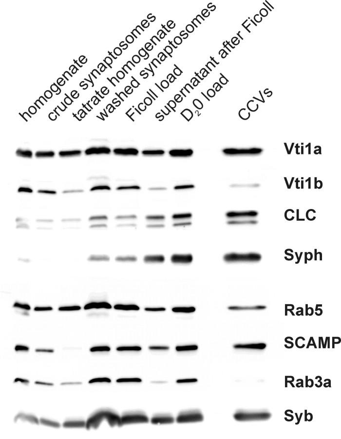
Specific soluble N-ethylmaleimide-sensitive factor attachment protein (SNAP) receptor (SNARE) proteins are required for different membrane transport steps. The SNARE Vti1a has been colocalized with Golgi markers and Vti1b with Golgi and the trans-Golgi network or endosomal markers in fibroblast cell lines. Here we study the distribution of Vti1a and Vti1b in brain. Vti1b was found in synaptic vesicles but was not enriched in this organelle. A brain-specific splice variant of Vti1a was identified that had an insertion of seven amino acid residues next to the putative SNARE-interacting helix. This Vti1a-beta was enriched in small synaptic vesicles and clathrin-coated vesicles isolated from nerve terminals. Vti1a-beta also copurified with the synaptic vesicle R-SNARE synaptobrevin during immunoisolation of synaptic vesicles and endosomes. Therefore, both synaptobrevin and Vti1a-beta are integral parts of synaptic vesicles throughout their life cycle. Vti1a-beta was part of a SNARE complex in nerve terminals, which bound N-ethylmaleimide-sensitive factor and alpha-SNAP. This SNARE complex was different from the exocytic SNARE complex because Vti1a-beta was not coimmunoprecipitated with syntaxin 1 or SNAP-25. These data suggest that Vti1a-beta does not function in exocytosis but in a separate SNARE complex in a membrane fusion step during recycling or biogenesis of synaptic vesicles.


Figures

Fig. 1.
Fig. 1.
Vti1a and Vti1b were expressed in all tissues. Homogenates were prepared from the indicated mouse tissues, and 20 μg separated by SDS-PAGE Western blots were stained with antisera against Vti1a or Vti1b. The Vti1a antiserum recognized a ubiquitous protein of 27 kDa and an additional slightly larger brain-specific band. Vti1b antiserum bound to a single band of 29 kDa.
Fig. 2.
Fig. 2.
The brain-specific splice variant Vti1a-β had an insertion of seven amino acid residues. A, A single band was PCR-amplified from a lung cDNA library, a double band from a cerebellum cDNA library using primers specific for Vti1a. The slightly larger band encoded Vti1a-β with a insertion of seven amino acids after Q114. B, Expression pattern of Vti1a and Vti1a-β in different tissues. RT-PCRs were performed using primer pairs amplifying both Vti1a and Vti1a-β (expected sizes, 511 and 490 bp;top), Vti1a only (forward primer annealing with codons 110–116, 265 bp; middle), or Vti1a-β only (forward primer annealing with Vti1a-β-specific codons 115–121, 253 bp;bottom). Vti1a was amplified from all tissues examined. Vti1a-β was amplified from the neuronal tissues cerebellum, cortex, and hippocampus, but not from lung, liver, kidney, or spleen.C, Alignment of Saccharomyces cerevisiaeVti1p, the C-terminal part of a predicted C. elegansprotein (GenBank accession number CAB16506), mouse Vti1b, rat Vti1a (GenBank accession number AF262221), and rat Vti1a-β (GenBank accession number AF262222). Filled circles indicate the beginning and end of the predicted SNARE-interacting helix (SNARE motif). The open circle marks the position of the conserved glutamine or aspartate residue in layer 0, andasterisks indicate hydrophobic positions in the heptade repeats.
Fig. 3.
Fig. 3.
Vti1a localized to the cell body as well as to nerve terminals of hippocampal neurons. Cultured hippocampal neurons were double stained for Vti1a or Vti1b and synaptobrevin. Vti1a and Vti1b were found in the cell body. In addition, Vti1a colocalized with synaptobrevin in nerve terminals. Scale bar, 20 μm.
Fig. 4.
Fig. 4.
Vti1a localized to the cell body as well as to mossy fiber terminals in hippocampal sections. Rat hippocampal sections were double stained for Vti1a or Vti1b and synaptobrevin. Vti1a and Vti1b were found in the cell bodies of pyramidal cells. Vti1a also colocalized with synaptobrevin in mossy fiber nerve terminals. Theinsets in the top panels show a threefold magnification of an area from the mossy fiber nerve terminals. Scale bar, 30 μm.
Fig. 5.
Fig. 5.
Vti1a-β copurified with synaptic vesicles. Synaptosomes (P2) were isolated from rat brain homogenate (H). The synaptosomes were osmotically lysed and separated into a low-speed membrane fraction (LP1) containing synaptic plasma membranes and a high-speed pellet (LP2) with synaptic vesicles. The synaptic vesicles were further purified by sucrose density gradient centrifugation and chromatography on a CPG column yielding the highly enriched fraction CPG3. Vti1a-β copurified with the synaptic vesicle marker synaptobrevin (Syb). Vti1b was present in synaptic vesicles but was not enriched compared with homogenate.
Fig. 6.
Fig. 6.
Vti1a was localized to synaptic vesicles using immunogold electron microscopy. The CPG3 fraction was adsorbed to coated grids and incubated with antisera against the synaptic vesicle marker synaptophysin (A), against Vti1a (B), or without antiserum (C) followed by 10 nm goat anti-rabbit IgG gold conjugates and negative staining. Scale bar, 500 nm.
Fig. 7.
Fig. 7.
Vti1a-β copurified with clathrin-coated vesicles isolated from nerve terminals. Clathrin-coated vesicles (CCVs) were isolated from synaptosomes with brain homogenate as the starting material. Highly enriched clathrin-coated vesicles were isolated from the high-speed pellet of lysed synaptosomes (Ficoll load) via the fractions Ficoll SN and D2O load. Vti1a-β copurified in parallel with the synaptic vesicle markers synaptobrevin (Syb), synaptophysin (Syp), and SCAMP in clathrin-coated vesicles. Clathrin light chain (CLC) was highly enriched. Very low amounts of Vti1b were found in clathrin-coated vesicles.
Fig. 8.
Fig. 8.
Vti1a-β was found on immunoisolated synaptic vesicles and endosomes. A 50,000 × g supernatant of rat brain homogenate was incubated with antibodies against synaptobrevin (Syb), rab3a, or rab5 coupled to Eupergit beads. Glycine was coupled to control beads. The bead fractions were washed and unbound, and bound fractions were separated by SDS-PAGE and analyzed by immunoblotting for different markers. Most Vti1a-β-containing organelles could be isolated with synaptobrevin and rab3a beads. A large fraction of Vti1a-β was found on rab5-containing organelles. Vti1b could also be immunoisolated with synaptobrevin, rab3a, and rab5 beads but to a smaller extend than Vti1a-β. Control beads did not bind any proteins, and the endoplasmic reticulum protein Sec61α was not bound to the immunobeads, indicating that the immunoisolations of organelles were specific.
Fig. 9.
Fig. 9.
Vti1a-β was not found in the exocytic SNARE complex. SNARE complexes were isolated from Triton X-100 extracts of synaptosomes using antisera against Vti1a, SNAP-25, syntaxin 1 (Syx1), or synaptobrevin (Syb). Vti1a antiserum did not coimmunoprecipitate syntaxin 1, SNAP-25, or synaptobrevin. SNAP-25, syntaxin 1, and synaptobrevin coimmunoprecipitated as a SNARE complex.
Fig. 10.
Fig. 10.
Vti1a-β was part of a SNARE complex that bound NSF and α-SNAP. Detergent extracts from lysed synaptosomes (high-speed membrane fraction LP2) were incubated without additions (−) or with NSF and α-SNAP and separated on a glycerol gradient. Fractions were analyzed by SDS-PAGE and immunoblotting. A subfraction of Vti1a-β and synaptobrevin (Syb) were shifted to denser fractions 11–13 in the presence of NSF and α-SNAP.
Fig. 11.
Fig. 11.
A C-terminal 14 kDa fragment of Vti1a-β with the SNARE motif was protease-protected under conditions of SNARE complex assembly but not disassembly. Detergent extracts from lysed synaptosomes (high-speed membrane fraction LP2) were incubated with NSF and α-SNAP plus ATP + Mg2+ (disassembly of SNARE complexes), ATPγS + Mg2+, or ATP + EDTA (inhibition of disassembly). SNARE complexes remained assembled without addition of NSF and α-SNAP. Fractions were incubated with trypsin and separated by SDS-PAGE. Immunoblots were developed with antisera against Vti1a (left), against the N-terminal half of Vti1a (middle), or against the C-terminal half of Vti1a (right).

References

    1. Advani RJ, Bae HR, Bock JB, Chao DS, Doung YC, Prekeris R, Yoo JS, Scheller RH. Seven novel mammalian SNARE proteins localize to distinct membrane compartments. J Biol Chem. 1998;273:10317–10324. - PubMed
    1. Advani RJ, Yang B, Prekeris R, Lee KC, Klumperman J, Scheller RH. VAMP-7 mediates vesicular transport from endosomes to lysosomes. J Cell Biol. 1999;146:765–775. - PMC - PubMed
    1. Alés E, Tabares L, Poyato JM, Valero V, Lindau M, de Toledo GA. High calcium concentrations shift the mode of exocytosis to the kiss-and-run mechanism. Nat Cell Biol. 1999;1:40–44. - PubMed
    1. Barnstable CJ, Hofstein R, Akagawa K. A marker of early amacrine cell development in rat retina. Dev Brain Res. 1985;20:286–290. - PubMed
    1. Bruns D, Engers S, Yang C, Ossig R, Jeromin A, Jahn R. Inhibition of transmitter release correlates with the proteolytic activity of tetanus toxin and botulinus toxin A in individual cultured synapses of Hirudo medicinalis. J Neurosci. 1997;17:1898–1910. - PMC - PubMed

Publication types

MeSH terms

LinkOut - more resources