. 2000 Aug 1;20(15):5748-55.
doi: 10.1523/JNEUROSCI.20-15-05748.2000.

Metabolic stress reversibly activates the Drosophila light-sensitive channels TRP and TRPL in vivo

Affiliations

Metabolic stress reversibly activates the Drosophila light-sensitive channels TRP and TRPL in vivo

K Agam et al. J Neurosci. .

Abstract

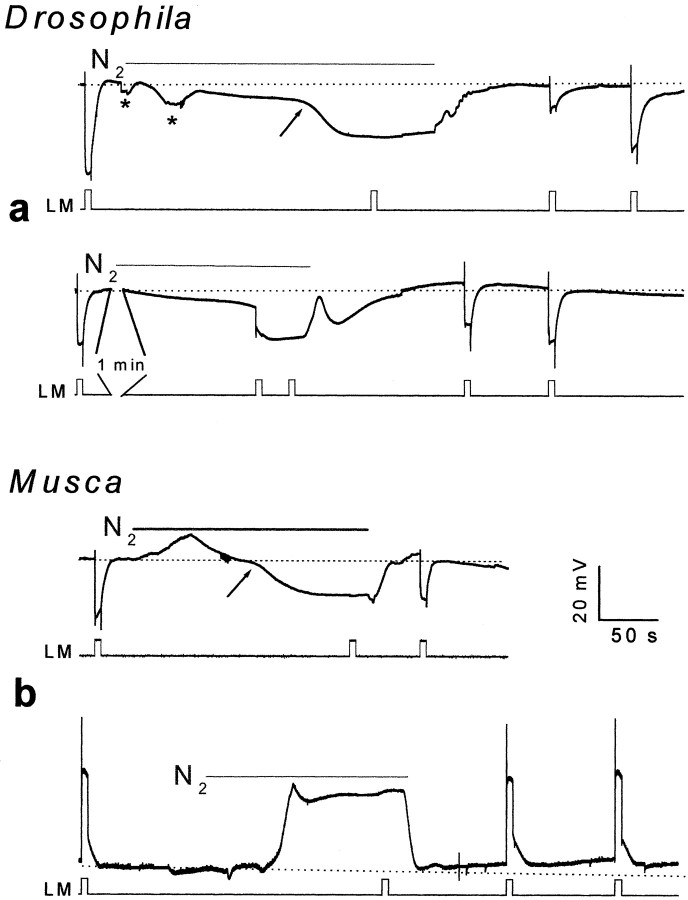
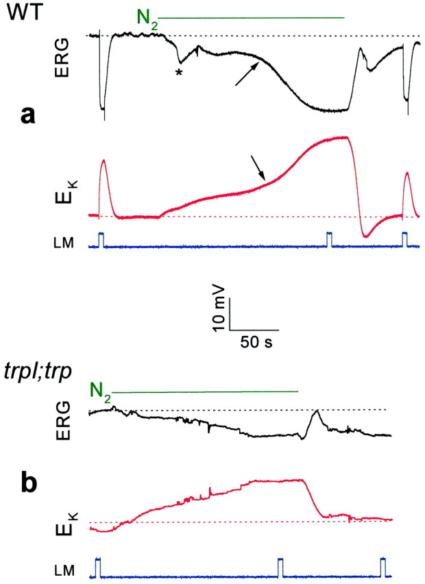
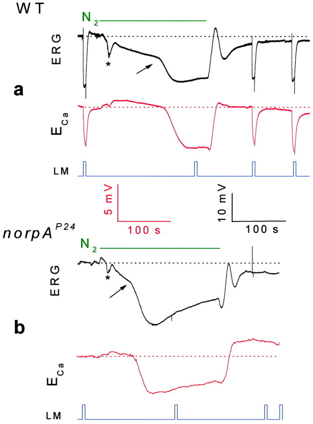
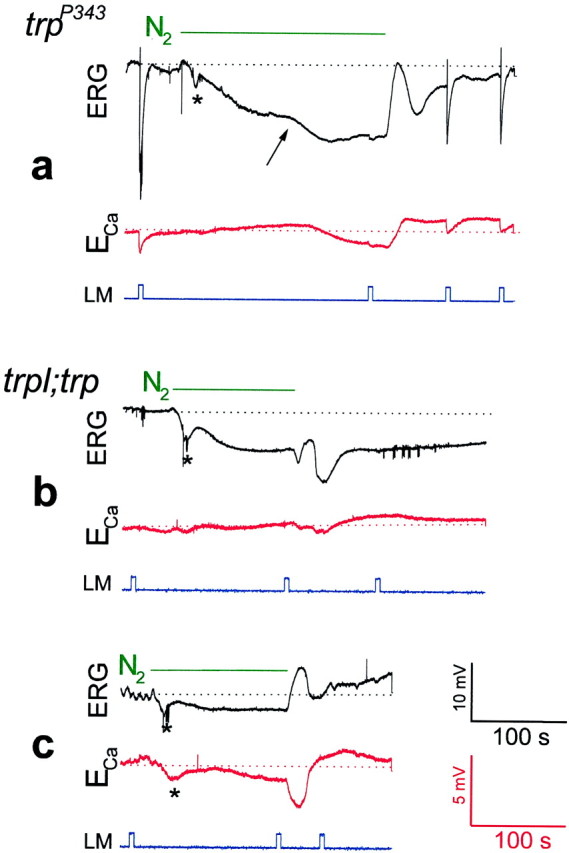
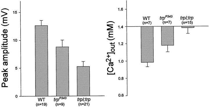
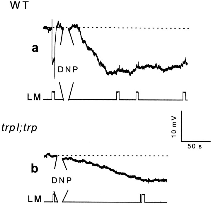
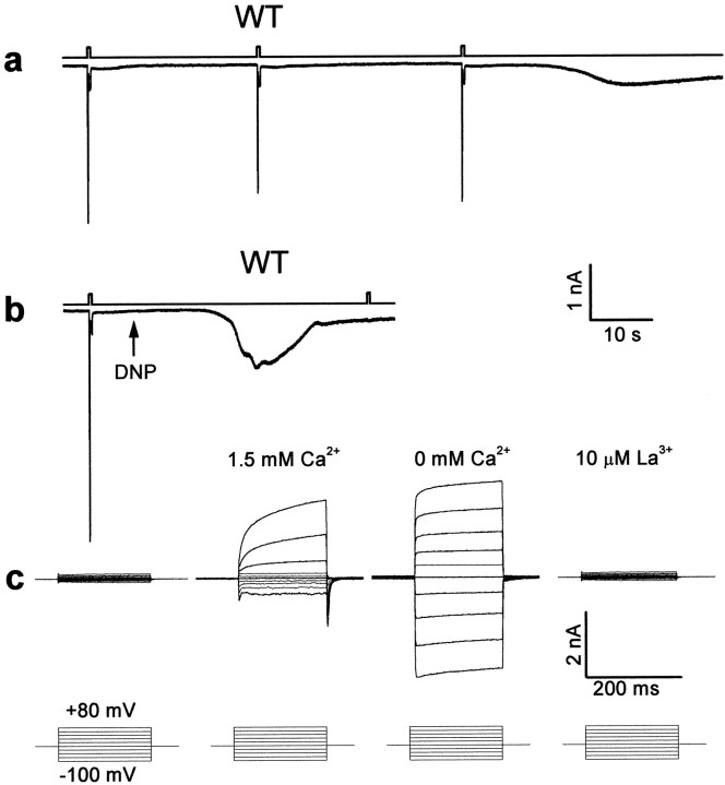
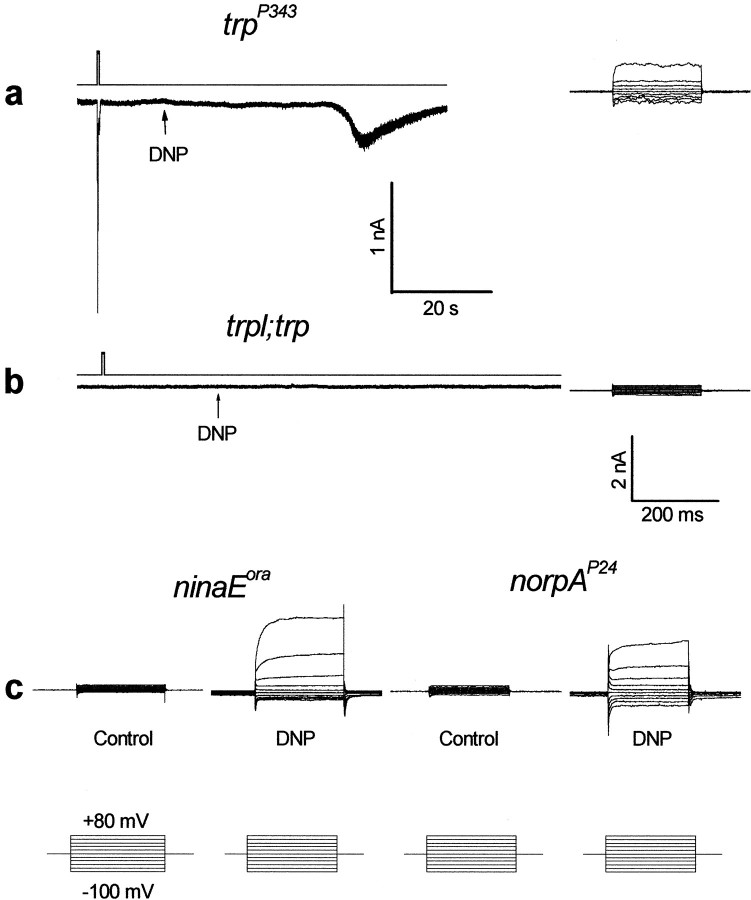
Drosophila transient receptor potential (TRP) is a prototypical member of a novel family of channel proteins underlying phosphoinositide-mediated Ca(2+) entry. Although the initial stages of this signaling cascade are well known, downstream events leading to the opening of the TRP channels are still obscure. In the present study we applied patch-clamp whole-cell recordings and measurements of Ca(2+) concentration by ion-selective microelectrodes in eyes of normal and mutant Drosophila to isolate the TRP and TRP-like (TRPL)-dependent currents. We report that anoxia rapidly and reversibly depolarizes the photoreceptors and induces Ca(2+) influx into these cells in the dark. We further show that openings of the light-sensitive channels, which mediate these effects, can be obtained by mitochondrial uncouplers or by depletion of ATP in photoreceptor cells, whereas the effects of illumination and all forms of metabolic stress were additive. Effects similar to those found in wild-type flies were also found in mutants with strong defects in rhodopsin, Gq-protein, or phospholipase C, thus indicating that the metabolic stress operates at a late stage of the phototransduction cascade. Genetic elimination of both TRP and TRPL channels prevented the effects of anoxia, mitochondrial uncouplers, and depletion of ATP, thus demonstrating that the TRP and TRPL channels are specific targets of metabolic stress. These results shed new light on the properties of the TRP and TRPL channels by showing that a constitutive ATP-dependent process is required to keep these channels closed in the dark, a requirement that would make them sensitive to metabolic stress.


Figures

Fig. 1.
Fig. 1.
Anoxia induced a rapid and reversible depolarization of fly photoreceptors in the dark and abolished light excitation. a, Top panel, Extracellular voltage change recordings of intact Drosophila eye in response to orange light (OG590, attenuated by 1 log unit, in all Figures) followed by application of anoxia (N2, as indicated) and by additional light pulses, which test the recovery from anoxia. The light monitor is indicated below (LM). Note that the second light pulse of the top panel and the third pulse at thebottom panel did not elicit any response. Thearrow indicates the onset of the second phase of the response to anoxia of larger amplitude. Movement artifacts, which probably resulted from movements of the fly eye after anoxia, are indicated by an asterisk (in all Figures).a, Bottom panel, The experiments of thetop traces were repeated in anotherDrosophila fly except that an additional orange light pulse was applied during the initial response to anoxia of small amplitude. A 1 min break is indicated in the bottom traces. b, Top panel, The experiments of a (top traces) were repeated in intact Musca fly. Bottom panel, Intracellular recordings from a single photoreceptor cell in response to the orange light pulses and anoxia. Note that the initial small and slow response to anoxia is absent.
Fig. 2.
Fig. 2.
Measurements of [K+]out in WT and thetrpl302;trp343mutant show that only the second and large phase of the response to anoxia arise from activation of TRP and TRPL channels.a, Extracellular voltage change (ERG,top panel, in black) and potentiometric measurements with a K+-selective microelectrode (EK, bottom panel, inred) in response to orange lights and anoxia, in wild-type (WT) Drosophila. On average, the maximal Δ[K+]out during anoxia was 7.26 ± 1.83 mm (n= 4), assuming that [K+]out in the dark is 4 mm (Sandler and Kirschfeld, 1991). The calibration applies for both the ERG and the potentiometric measurements with the K+-selective microelectrode.b, The experiments of a were repeated in the double mutanttrpl302;trp343. Note that there is no response to light, and the second phase of the response to anoxia is absent. On average, the maximal Δ[K+]out was 2.64 ± 0.54 mm (n = 4) during anoxia.
Fig. 3.
Fig. 3.
Anoxia activated the TRP and TRPL channels in both WT (a) and the PLC null mutant (norpAP24, b) as monitored by Ca2+ influx. Extracellular voltage change (ERG, top traces in botha and b, in black) and potentiometric measurements with Ca2+-selective microelectrode (ECa, bottom traces in both a and b, inred) in response to orange lights and anoxia in WTDrosophila andnorpAP24 mutant are shown. Note that there is no response to light in thenorpAP24 mutant and the initial slow phase of the electrical response to anoxia is missing in the Ca2+ signals of both WT and the mutant. The calibrations for the ERG records are indicated in black, and the calibrations for the potentiometric measurements with the Ca2+-selective microelectrode are indicated inred (in Figs. 3 and 4).
Fig. 4.
Fig. 4.
Genetic elimination of TRP (a) resulted in a reduced Ca2+ influx through the remaining TRPL channels in response to anoxia, whereas elimination of both TRP and TRPL (b, c) virtually abolished TRP- and TRPL-dependent signals and Ca2+ influx. The same paradigm of Figure 3 was repeated in Figure 4 except that the null trp mutanttrpP343 (a) and the null double mutanttrpl302;trpP343(in two different flies) (b, c) were used. Note that in the trpP343mutant the second phase of the electrical response to anoxia and the Ca2+ influx were relatively small but were maintained as long as anoxia was applied, in contrast to the transient responses to light. Also note that there is no response to light in the trpl;trp mutant and that the second phase of the electrical response to anoxia was absent. The negative small and slow Ca2+ signal in c revealed variability in sign and appeared in only ∼30% of the mutant flies.
Fig. 5.
Fig. 5.
Histograms showing maximal voltage changes, which include both the slow and fast phases (left) and Δ[Ca2+]out (right), in response to anoxia in WT, trp, andtrpl;trp mutants. The error bars are SEM.
Fig. 6.
Fig. 6.
The mitochondria uncoupler 2,4-dinitrophenol (DNP) mimicked the effects of anoxia as monitored by extracellular voltage changes. a, The paradigm of Figure1a was repeated in WT Drosophila, except that an additional pipette containing either 1 or 10 mm DNP in Ringer's solution was inserted into the retina, and DNP was applied by pressure injection (during the 30 sec break in a andb). It is estimated that DNP is diluted ∼10- to 40-fold in the eye. Control injections of Ringer's solution without DNP had no effect. b, The paradigm of awas repeated in the double mutanttrpl302;trpP343.
Fig. 7.
Fig. 7.
Single-cell functional analysis by whole-cell recordings from newly eclosed flies showing that depletion of ATP activates the TRP and TRPL channels of WT cells. Omission of ATP and NAD from the recording pipette (in all traces) induced, after a few light pulses, or after application of DNP, a constitutive activation of the light-sensitive channels. This channel activity was indicated by the appearance of a transient phase, which was followed by a sustained noisy inward current that had all the characteristics of the TRP- or TRPL-dependent current. None of these currents were observed in the presence of ATP and NAD in the pipette or before the dark inward current was induced. This was demonstrated by application of voltage steps in the dark during whole-cell recordings from photoreceptor cells, which revealed only small leak currents (c,left). The establishment of the whole-cell recordings took place ∼10 sec before the beginning of the traces in this Figure.a, Typical light-induced currents of a WT cell in response to three orange lights was followed by the appearance of slow inward current in the dark when the pipette solution had no ATP and NAD. The membrane voltage was held at −50 mV. The top traces in each pair indicate the duration of the orange light stimuli. b, The onset of the dark inward current was accelerated by application of 0.1 mm DNP (arrow, in a different cell). The inward current was induced 19.7 ± 3.5 sec after application of DNP (n = 4) compared with 108.5 ± 19.7 sec (n = 8) without application of DNP under similar recording conditions (a). Note that no additional response to light was obtained after the dark inward current was induced. c, A comparison of families of current traces elicited by a series of voltage steps in the range of −100 to +80 mV in steps of 20 mV (bottom row), from photoreceptors of wild-type flies under the following conditions: in the dark before the inward current was induced, at the peak of the inward current at 1.5 mm external Ca2+, after Ca2+ was removed from the external medium (0 Ca2+), and after 10 μmLa3+ was applied to the external medium (as indicated).
Fig. 8.
Fig. 8.
The effects of DNP on Drosophilamutants. a, A relatively slow induction of a small and noisy inward current in the dark was observed intrpP343 after application of 0.1 mm DNP (arrow) during recordings without ATP and NAD in the pipette. Inward currents could not be induced without application of DNP under similar recording conditions. The right traces show a family of current traces elicited by a series of voltage steps as in Figure 7c at 1.5 mmexternal Ca2+. Similar families of current traces of similar amplitudes were observed at 0 Ca2+ medium (n = 3). b, Recording from thetrpl302;trpP343double mutant using pipettes in which ATP and NAD were omitted and 0.1 mm DNP was applied to the external medium as indicated. No dark inward current was observed even 16 min after the beginning of whole-cell recordings. The right traces show a family of current traces elicited by a series of voltage steps as in Figure7c at 1.5 mm external Ca2+. Similar results were obtained from four different cells in which DNP was included in the recording pipette. Note that neither light-induced currents nor inward currents in the dark could be induced in the double mutant. c, The effects of DNP (applied through the pipette) on theninaEora, q1, andnorpAP24 mutants. Thetraces are families of current traces elicited by a series of voltage steps as in Figure 7c at 1.5 mm external Ca2+. The typical TRP-dependent current is observed only in cells in which the recording pipette included 0.1 mm DNP but not in control cells of the same retinae, as indicated. Similar results were obtained from three to six different cells of each mutant. The traces recorded from theq1 mutant are not shown. The lower calibration applies to all families of current traces elicited by a series of voltage steps.

References

    1. Arslan P, Corps AN, Hesketh TR, Metcalfe JC, Pozzan T. cis-Unsaturated fatty acids uncouple mitochondria and stimulate glycolysis in intact lymphocytes. Biochem J. 1984;217:419–425. - PMC - PubMed
    1. Balzer M, Lintschinger B, Groschner K. Evidence for a role of Trp proteins in the oxidative stress-induced membrane conductances of porcine aortic endothelial cells. Cardiovasc Res. 1999;42:543–549. - PubMed
    1. Baumann F, Mauro A. Effect of hypoxia on the change in membrane conductance evoked by illumination in arthropod photoreceptors. Nat New Biol. 1973;244:146–148. - PubMed
    1. Berridge MJ. Capacitive calcium entry. Biochem J. 1995;312:1–11. - PMC - PubMed
    1. Berridge MJ, Lipp P, Bootman MD. Signal transduction: the calcium entry pas de deux. Science. 2000;287:1604–1605. - PubMed

Publication types

MeSH terms