. 2000 Aug 1;20(15):5775-81.
doi: 10.1523/JNEUROSCI.20-15-05775.2000.

BDNF protects the neonatal brain from hypoxic-ischemic injury in vivo via the ERK pathway

Affiliations

BDNF protects the neonatal brain from hypoxic-ischemic injury in vivo via the ERK pathway

B H Han et al. J Neurosci. .

Abstract

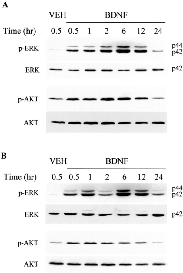
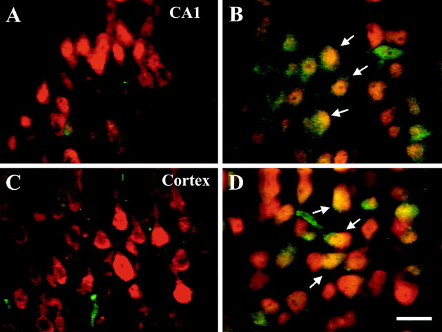
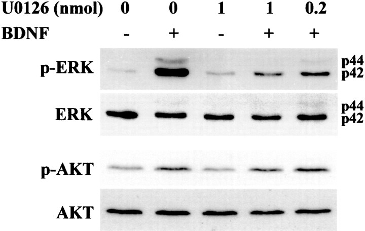
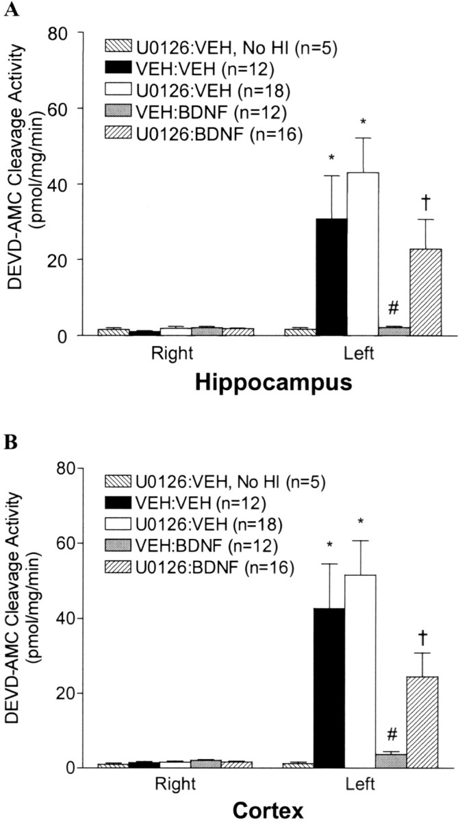
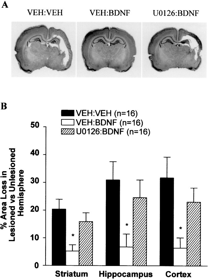
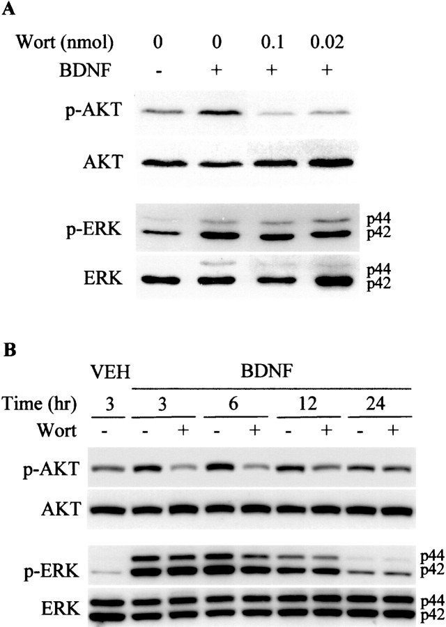
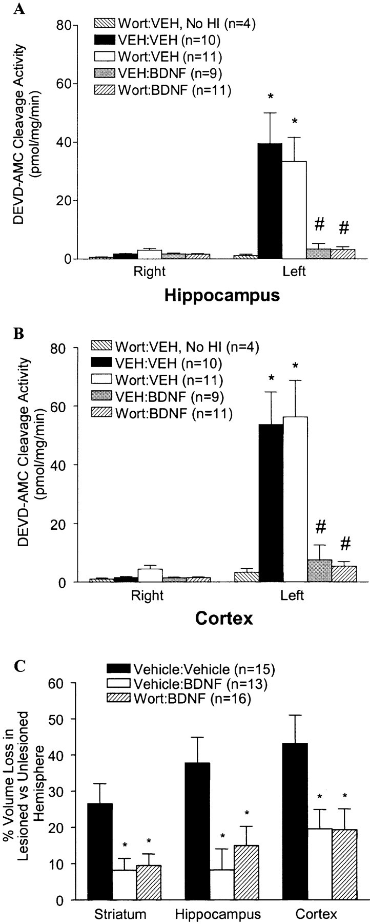
Neurotrophins activate several different intracellular signaling pathways that in some way exert neuroprotective effects. In vitro studies of sympathetic and cerebellar granule neurons have demonstrated that the survival effects of neurotrophins can be mediated via activation of the phosphatidylinositol 3-kinase (PI3-kinase) pathway. Neurotrophin-mediated protection of other neuronal types in vitro can be mediated via the extracellular signal-related protein kinase (ERK) pathway. Whether either of these pathways contributes to the neuroprotective effects of neurotrophins in the brain in vivo has not been determined. Brain-derived neurotrophic factor (BDNF) is markedly neuroprotective against neonatal hypoxic-ischemic (H-I) brain injury in vivo. We assessed the role of the ERK and PI3-kinase pathways in neonatal H-I brain injury in the presence and absence of BDNF. Intracerebroventricular administration of BDNF to postnatal day 7 rats resulted in phosphorylation of ERK1/2 and the PI3-kinase substrate AKT within minutes. Effects were greater on ERK activation and occurred in neurons. Pharmacological inhibition of ERK, but not the PI3-kinase pathway, inhibited the ability of BDNF to block H-I-induced caspase-3 activation and tissue loss. These findings suggest that neuronal ERK activation in the neonatal brain mediates neuroprotection against H-I brain injury, a model of cerebral palsy.


Figures

Fig. 1.
Fig. 1.
BDNF increases phosphorylation of ERK1/2 and AKT in the neonatal brain. P7 rat pups received an ICV injection of vehicle (PBS) or BDNF (5 μg in PBS). Brain tissues from the hippocampus (A) and cortex (B) were dissected at the various times indicated and lysed. Proteins were separated by 12.5% SDS-PAGE and transferred to nitrocellulose membranes. Immunoblotting was first performed with an anti-phospho-ERK1/2 antibody; blots were stripped and reprobed with anti-phospho-AKT, anti-ERK1/2, and anti-AKT antibodies. On theright in A and B,p44 corresponds to ERK1, and p42corresponds to ERK2. Similar results were found in four independent experiments.
Fig. 2.
Fig. 2.
BDNF induces phosphorylation of ERK1/2 in neurons. Brain sections were prepared from P7 rats 30 min after an ICV injection of either vehicle (A, C) or BDNF (B, D) (n = 3/group). Brain sections from the hippocampus (A, B) and cortex (C, D) were labeled with anti-neuronal nucleus antibody NeuN (red) and anti-phospho-ERK1/2 antibody specific to the phosphorylated form of ERK1/2 (green). Arrows inB and D indicate yellowcells in which NeuN and p-ERK1/2 are colocalized. Scale bar, 20 μm.
Fig. 3.
Fig. 3.
U0126 blocks BDNF-mediated ERK1/2 phosphorylationin vivo. P7 rats were ICV injected with vehicle (1 μl of DMSO in 4 μl of PBS) or U0126 (0.2 or 1 nmol/animal) 20 min before receiving an ICV injection of vehicle or BDNF (5 μg/animal) as indicated. Cortical tissues prepared 1 hr after the injection were lysed, and proteins (50 μg/lane) were separated by SDS-PAGE and subjected to immunoblotting with an anti-phospho-ERK1/2 antibody specific to phosphorylated ERK1/2. Blots were stripped and subsequently reprobed with anti-ERK, anti-phospho-AKT, and anti-AKT. Data shown are representative of six independent experiments.
Fig. 4.
Fig. 4.
Inhibition of the ERK1/2 pathway blocks BDNF-mediated inhibition of caspase-3-like activity. P7 rats received ICV injections of vehicle or U0126 (1 nmol/animal) followed 15 min later by an ICV injection of vehicle or BDNF (5 μg/animal) just before the carotid ligation and exposure to 2.5 hr of hypoxia. Twenty-four hours after H-I, brain tissues from the hippocampus (A) and cortex (B) contralateral (right) and ipsilateral (left) to the ligation were dissected and subjected to DEVD-AMC cleavage assay as described in Materials and Methods. Note that there is no activation of caspase-3-like activity in the group treated with U0126 without subsequent H-I (No HI). Data represent the mean ± SEM; *p < 0.05 compared with the contralateral (right) hemisphere, #p < 0.05 comparing ipsilateral hemispheres of VEH:BDNF with that of the VEH:VEH group, and †p < 0.05 comparing ipsilateral hemispheres from theU0126:BDNF group with that of theVEH:BDNF group. Data were analyzed by ANOVA and Dunn's multiple comparison method.
Fig. 5.
Fig. 5.
Inhibition of the ERK1/2 pathway blocks BDNF-mediated neuroprotection against neonatal H-I. P7 rats were treated with VEH:VEH, VEH:BDNF, orU0126:BDNF before the carotid ligation and exposure to hypoxia for 2.5 hr. One week later, animals were killed, and brain sections were stained with cresyl violet. A, Examples of the degree of injury of representative animals from each treatment group are shown. Note the marked unilateral hemispheric tissue loss in the cortex and hippocampus in VEH:VEH andU0126:BDNF animals, whereas there is little to no tissue loss in the VEH:BDNF animal. B,BDNF-mediated neuroprotection was significantly inhibited by U0126. Regional area loss from the striatum, hippocampus, and cortex of each group was assessed as described in Materials and Methods. Data represent the mean ± SEM; *p < 0.05 comparing VEH:BDNF with either theVEH:VEH or the U0126:BDNF group. Data were analyzed by ANOVA and Dunn's multiple comparison method.
Fig. 6.
Fig. 6.
BDNF-mediated AKT but not ERK1/2 phosphorylation is blocked by wortmannin in vivo. P7 rats received ICV injections of vehicle (1 μl of DMSO in 4 μl of PBS) or wortmannin (Wort; 0.1 or 0.02 nmol/animal in A; 0.1 nmol/animal in B) followed 15 min later by an ICV injection of vehicle or BDNF (5 μg/animal) as indicated. Cortical tissues prepared 1 hr after the injection in A or at later time points in B were lysed, and proteins (50 μg/lane) were separated by SDS-PAGE and subjected to immunoblotting with an anti-phospho-AKT antibody specific to phosphorylated AKT. Blots were stripped and subsequently reprobed with anti-phospho-ERK1/2, anti-ERK, and anti-AKT. Data shown are representative of four independent experiments.
Fig. 7.
Fig. 7.
Inhibition of the AKT pathway does not block BDNF-mediated neuroprotection against neonatal H-I. A, B, P7 rats received ICV injections of VEH or wortmannin (0.1 nmol/animal) followed 15 min later by ICV injections of VEH or BDNF (5 μg/animal) before the carotid ligation and exposure to hypoxia for 2.5 hr. Twenty-four hours after H-I, brain tissues from the hippocampus (A) and cortex (B) contralateral (right) and ipsilateral (left) to the ligation were dissected and subjected to DEVD-AMC cleavage assay as described in Materials and Methods. Note that there is no activation of caspase-3-like activity in the group treated with wortmannin without subsequent H-I. Data represent the mean ± SEM; *p < 0.05 comparing VEH:VEH and Wort:VEHipsilateral with contralateral hemispheres; #p < 0.05 comparing ipsilateral hemispheres of VEH:BDNF andWort:BDNF with ipsilateral hemispheres of eitherVEH:VEH or Wort:VEH groups. Data were analyzed by ANOVA and Dunn's multiple comparison. C, P7 rats were treated with Vehicle:Vehicle,Vehicle:BDNF, or Wort:BDNF before the carotid ligation and exposure to hypoxia for 2.5 hr. One week later, animals were killed, and brain sections were stained with cresyl violet. The regional area loss from the striatum, hippocampus, and cortex of each group was assessed as described in Materials and Methods. Data represent the mean ± SEM; *p < 0.05 comparing Vehicle:Vehicle with either theVehicle:BDNF or the Wort:BDNF group. Data were analyzed by ANOVA and Dunn's multiple comparison method.

References

    1. Alessandro A, Namura S, Moskowitz MA, Bonventre JV. MEK1 protein kinase inhibition protects against damage resulting from focal cerebral ischemia. Proc Natl Acad Sci USA. 1999;96:12866–12869. - PMC - PubMed
    1. Blenis J. Signal transduction via the MAP kinases: proceed at your own Rsk. Proc Natl Acad Sci USA. 1993;90:5889–5892. - PMC - PubMed
    1. Bonni A, Brunet A, West AE, Datta SR, Takasu MA, Greenberg ME. Cell survival promoted by the Ras-MAPK signaling pathway by transcription-dependent and -independent mechanisms. Science. 1999;286:1358–1365. - PubMed
    1. Chao MV. Neurotrophin receptors: a window into neuronal differentiation. Neuron. 1992;9:583–593. - PubMed
    1. Chen KS, Nishimura MC, Armanini MP, Crowley C, Spencer SD, Phillips HS. Disruption of a single allele of the nerve growth factor gene results in atrophy of basal forebrain cholinergic neurons and memory deficits. J Neurosci. 1997;17:7288–7296. - PMC - PubMed

Publication types

MeSH terms