. 2000 Aug 1;20(15):5782-91.
doi: 10.1523/JNEUROSCI.20-15-05782.2000.

Depletion of a microtubule-associated motor protein induces the loss of dendritic identity

Affiliations

Depletion of a microtubule-associated motor protein induces the loss of dendritic identity

W Yu et al. J Neurosci. .

Abstract

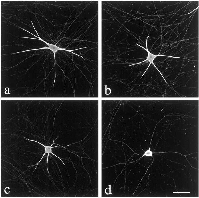
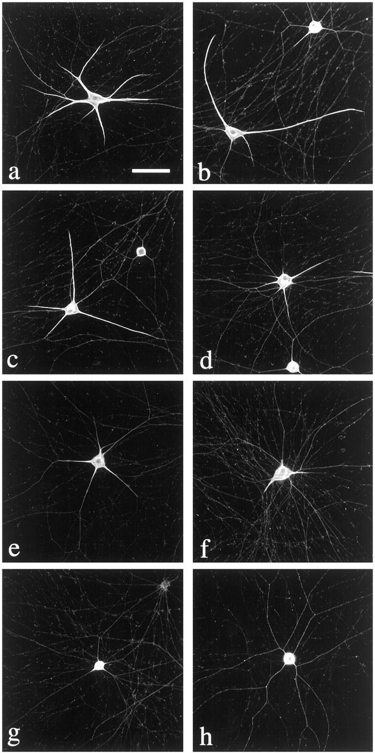
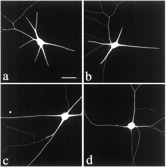
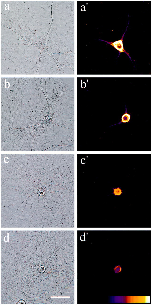
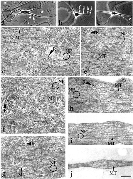
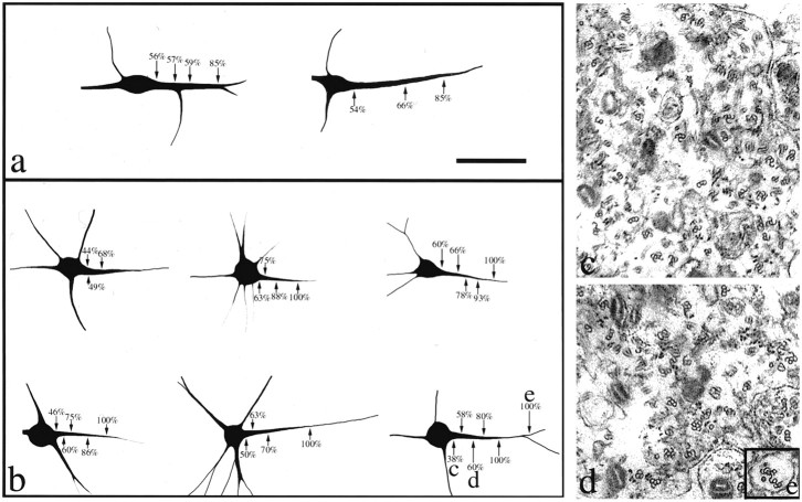
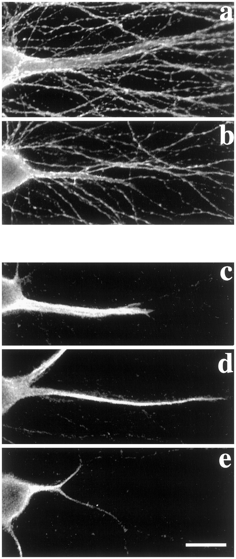
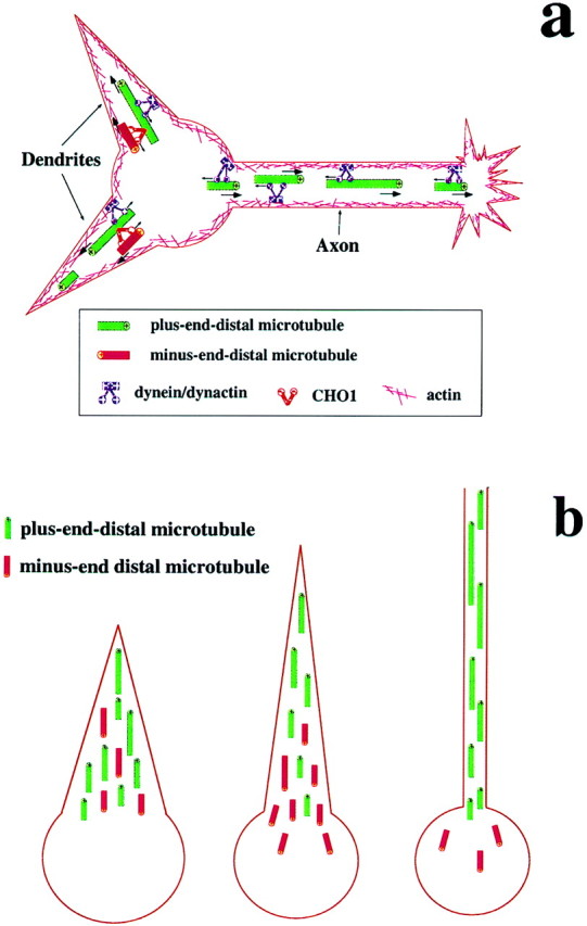
Dendrites are short stout tapering processes that are rich in ribosomes and Golgi elements, whereas axons are long thin processes of uniform diameter that are deficient in these organelles. It has been hypothesized that the unique morphological and compositional features of axons and dendrites result from their distinct patterns of microtubule polarity orientation. The microtubules within axons are uniformly oriented with their plus ends distal to the cell body, whereas microtubules within dendrites are nonuniformly oriented. The minus-end-distal microtubules are thought to arise via their specific transport into dendrites by the motor protein known as CHO1/MKLP1. According to this model, CHO1/MKLP1 transports microtubules with their minus ends leading into dendrites by generating forces against the plus-end-distal microtubules, thus creating drag on the plus-end-distal microtubules. Here we show that depletion of CHO1/MKLP1 from cultured neurons causes a rapid redistribution of microtubules within dendrites such that minus-end-distal microtubules are chased back to the cell body while plus-end-distal microtubules are redistributed forward. The dendrite grows significantly longer and thinner, loses its taper, and acquires a progressively more axon-like organelle composition. These results suggest that the forces generated by CHO1/MKLP1 are necessary for maintaining the minus-end-distal microtubules in the dendrite, for antagonizing the anterograde transport of the plus-end-distal microtubules, and for sustaining a pattern of microtubule organization necessary for the maintenance of dendritic morphology and composition. Thus, we would conclude that dendritic identity is dependent on forces generated by CHO1/MKLP1.


Figures

Fig. 1.
Fig. 1.
Morphology of cultured rat sympathetic neurons revealed by neurofilament immunostaining. Shown here are examples of the morphology of rat sympathetic neurons that were cultured in the presence of OP-1 to promote robust dendritic differentiation. After 2 weeks in culture, the vast majority of the neurons showed thick, tapering dendrites. Cultures were immunostained with an antibody to a poorly phosphorylated neurofilament epitope that is highly enriched in dendrites compared to axons. Most of the neurons showed three or more robust thick, tapering curvaceous dendrites that were at least 30–50 μm in length. Some neurons showed a somewhat less robust dendritic arbor, but clearly showed unmistakable dendrites. Fewer than 10% of the neurons showed no robust dendrites (d). Scale bar, 30 μm.
Fig. 2.
Fig. 2.
Morphology of cultured rat sympathetic neurons exposed to CHO1/MKLP1-antisense for 1 d revealed by neurofilament immunostaining. Shown here are neurons treated with sense or antisense oligonucleotides specific for CHO1/MKLP1. The neurons shown here were immunostained with the same neurofilament antibody as in Figure 1. a shows a sense control with a robust dendritic arbor similar to that observed in typical control neurons. The remaining panels show antisense-treated neurons with clearly altered morphologies. The neuron shown in b displays dendrites that are longer than typical control dendrites, less tapered but still curvaceous. The vast majority of the experimental dendrites lost their curvaceous appearance and appeared straight and taut.c shows a neuron with dendrites that are somewhat thinner and less tapered than control dendrites, and less curvaceous. At least one of the dendrites is longer than typical controls. The remaining panels (d–h) show neurons in more advanced stages of their loss of dendritic morphology. The tapered regions of the dendrites appear to wither in a distoproximal fashion, but the actual length of the processes is difficult to assess because of bundling with neighboring axons. Scale bar, 40 μm.
Fig. 3.
Fig. 3.
Morphology of individual neurons revealed by Lucifer yellow dye injections. Immunostains shown in Figures 1 and 2suggest that during antisense treatment, the tapering regions of the dendrites wither, but it is unclear whether the entire length of the process increases or decreases because of the bundling of the dendrites with axons from neighboring cells. To investigate this issue, we injected Lucifer yellow into individual neurons to reveal their neuritic arbor. a shows an untreated neuron,b shows a sense-treated neuron, and c andd show antisense-treated neurons. Treatments were for 1 d. In the untreated and sense-treated neurons, the tips of the dendrites are clearly distinguishable. Typical dendrites were 30–50 μm in length. In the antisense-treated neurons, the dendrites are substantially longer than controls. In many cases, the dendrites had elongated to well over 100 μm over the first day in antisense. Scale bar, 30 μm.
Fig. 4.
Fig. 4.
Immunofluorescence analyses on the levels of CHO1/MKLP1 in control and antisense-treated neurons. After 1 d in antisense, cultures were fixed in cold methanol, and CHO1/MKLP1 was visualized using immunofluorescence microscopy. DIC images of the fixed cells are shown in the left-hand column, whereas corresponding immunofluorescence images are shown in theright-hand column. a anda′ show an untreated neuron with robust dendrites and strong CHO1/MKLP1 immunoreactivity in the cell body and dendrites but not the axonal network. b and b′ show another untreated neuron with somewhat less robust dendrites. The immunoreactivity is correspondingly somewhat less intense.c and c′ show an antisense-treated neuron wherein the withered tapered regions of dendrites are still apparent. Immunoreactivity is significantly lower than in control neurons.d and d′ show an antisense-treated neuron wherein the dendrites have almost completely reverted to an axonal morphology. The immunoreactivity is even lower yet. Scale bar, 35 μm.
Fig. 5.
Fig. 5.
Electron microscopic analyses on dendrites of control and CHO1/MKLP1-antisense-treated neurons. Top panels show phase-contrast micrographs of embedded samples.a is an untreated neuron, whereas b andc are antisense-treated. The remaining panels show electron micrographs from the indicated regions. MT, Microtubule; NF, neurofilament bundles typical of dendrites; R, ribosomes. Control neurons show abundant ribosomes near the cell body and fewer but still abundant levels farther down the length of the dendrite. Microtubules are scattered near the cell body, but more paraxial (but still not as paraxial as in axons) farther down the length of the dendrite. Neurofilament bundles are plentiful throughout. Ribosomes are still plentiful near the cell body of antisense-treated dendrites, but are dramatically diminished with distance, and virtually absent from the thinner distal regions of the original dendrite and the newly grown regions. Neurofilament bundles remain plentiful throughout most of the original length of the dendrite, but only sparse unbundled neurofilaments are present in the newly grown regions. Microtubules appear at relatively lower levels within the more proximal regions of the original dendrite, but at higher levels more distally. The microtubules appear to be more paraxial than in controls especially in the more distal regions of the antisense-treated dendrites. The levels of internal membranous elements (presumably Golgi) also appear to diminish with antisense treatment. Thus, the cytoplasm of the antisense-treated dendrites becomes more axonal in character. The change in microtubule distribution is consistent with the idea that the minus-end-distal microtubules are moving out of the dendrite back toward the cell body, and the plus-end-distal microtubules are translocating anterogradely during antisense treatment. Scale bar, 0.5 μm.
Fig. 6.
Fig. 6.
Microtubule polarity analyses on control and CHO1/MKLP1-antisense treated cultures. The standard “hooking” procedure was used to assess microtubule polarity orientation. Clockwise hooks indicate plus-end-distal microtubules, whereas counterclockwise hooks indicate minus-end-distal microtubules.a shows the data from two control dendrites, whereasb shows the data from six antisense-treated dendrites. The data are expressed as the percentages of clockwise hooks in different sampled regions. In control dendrites, slightly more than half of the hooks were clockwise in proximal and middle regions, with progressively higher percentages in the more distal regions. In dendrites that had completely thinned along their lengths, the percentage of clockwise hooks was >95%, indicating uniformly plus-end-distal microtubules (data not shown). The dendrites shown inb are “transitional,” in the sense that they still showed withering tapered regions. In all cases, the thinner distal regions showed predominantly or entirely clockwise hooks. In two of the six cases, the proportion of plus-end-distal microtubules was significantly higher than controls at corresponding sites throughout the length of the dendrite. In four of the six cases, this increase in the proportion of plus-end-distal microtubules was observed throughout most of the length of the dendrite, except in the most proximal region near the cell body, which actually showed a reduction in the proportion of clockwise hooks. Examples of the electron micrographs are shown inc–e, and the corresponding regions of the dendrite from which the electron micrographs were taken are shown bylettered-marked arrows in b. Scale bar:a, b, 40 μm; c–e, 0.45 μm.
Fig. 7.
Fig. 7.
Tau-1 and MAP-2 immunofluorescence analyses on control and CHO1/MKLP1 antisense-treated neurons. aand b show an untreated and an antisense-treated neuron, both stained with the tau-1 antibody. The dendrites of untreated cultures stained lightly and were clearly surrounded by more intensely-staining axons (a). The withering regions of the antisense-treated dendrites showed no apparent increase in staining for tau-1 and were still surrounded by more intensely stained axons (b). a shows an untreated neuron, whereas d and e show antisense-treated neurons, all stained for full-length MAP-2. The dendrites of untreated cultures stained intensely for full-length MAP-2, whereas the axons showed little or no staining (c). As the dendrites thinned and elongated in response to antisense-treatment, there was no immediate diminution in MAP-2 staining (d), although the intense MAP-2 staining was gradually diminished as the thick tapering regions of the dendrites became progressively more withered (e). Scale bar, 12 μm.
Fig. 8.
Fig. 8.
A model for the role of CHO1/MKLP1 in the dendrite. In our model (a), cytoplasmic dynein transports microtubules with their plus ends leading (pushing against the actin cytomatrix) into the axon and the dendrites, whereas CHO1/MKLP1 transports microtubules anterogradely with their minus-ends-leading (pushing against the plus-end-distal microtubules) into the dendrites but not the axon. The forces generated by CHO1/MKLP1 create drag on the plus-end-distal microtubules, which impedes their dynein-driven transport. Both the suppression of dynein-driven microtubule transport and the nonuniform microtubule polarity pattern itself result in the characteristic morphological and compositional features that distinguish dendrites from the axon. This model makes certain predictions regarding what would happen if CHO1/MKLP1 were depleted (b). The model predicts that when CHO1/MKLP1 is depleted, the forces generated by cytoplasmic dynein should no longer be antagonized. The plus-end-distal microtubules should therefore be better able to move anterogradely, and the minus-end-distal microtubules should be transported (with their plus ends leading) back toward the cell body. As the minus-end-distal microtubules are cleared and the plus-end-distal microtubules move rapidly forward, the dendrite should gradually lose its taper, thin out, and elongate. These predictions are consistent with the redistribution of microtubules and the alterations in dendritic morphology observed in our depletion studies. Observed compositional changes are also consistent with the predictions of the model (data not shown in the schematic).

References

    1. Ahmad FJ, Echeverri CJ, Vallee RB, Baas PW. Cytoplasmic dynein and dynactin are required for the transport of microtubules into the axon. J Cell Biol. 1998;140:246–256. - PMC - PubMed
    1. Ahmad FJ, Hughey J, Wittmann T, Hyman A, Greaser M, Baas PW. Motor proteins regulate force interactions between microtubules and microfilaments in the axon. Nat Cell Biol. 2000;2:276–280. - PubMed
    1. Baas PW. Microtubules and neuronal polarity: lessons from mitosis. Neuron. 1999;22:23–31. - PubMed
    1. Baas PW. Strategies for studying microtubule transport in the neuron. Microsc Res Tech. 2000;48:75–84. - PubMed
    1. Baas PW, Ahmad FJ. The transport properties of axonal microtubules establish their polarity orientation. J Cell Biol. 1993;120:1427–1437. - PMC - PubMed

Publication types

MeSH terms

LinkOut - more resources