. 2000 Aug 1;20(15):5792-801.
doi: 10.1523/JNEUROSCI.20-15-05792.2000.

Netrin-1 promotes thalamic axon growth and is required for proper development of the thalamocortical projection

Affiliations

Netrin-1 promotes thalamic axon growth and is required for proper development of the thalamocortical projection

J E Braisted et al. J Neurosci. .

Abstract

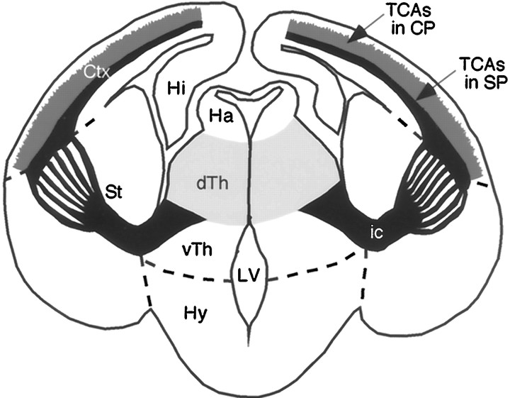
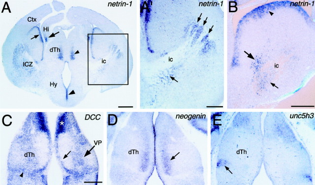
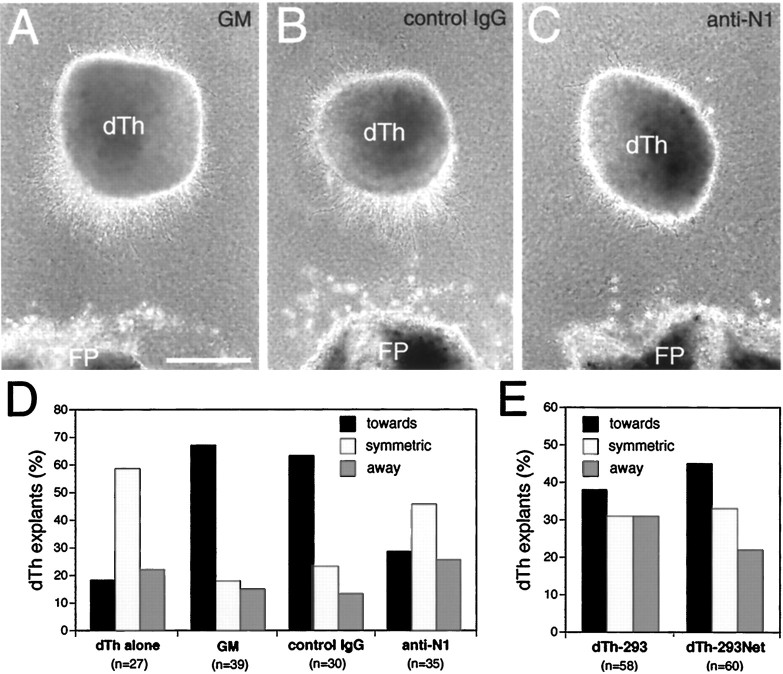
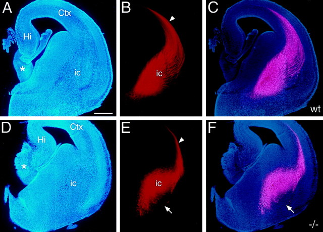
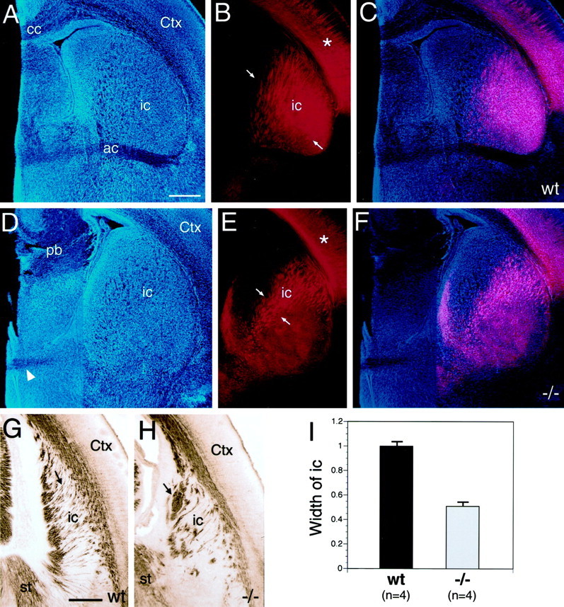
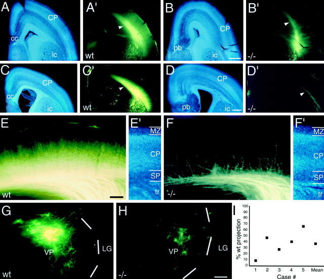
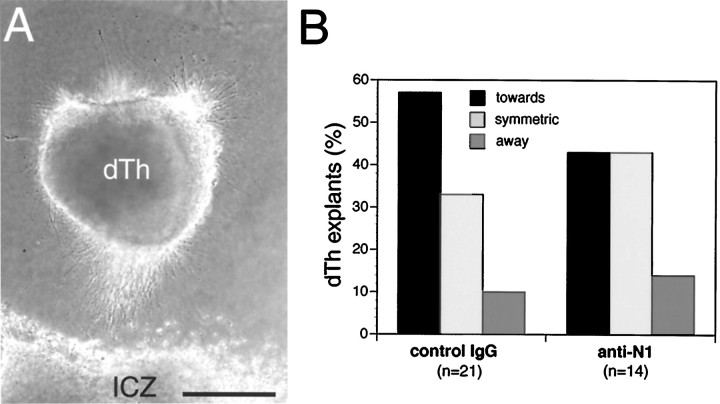
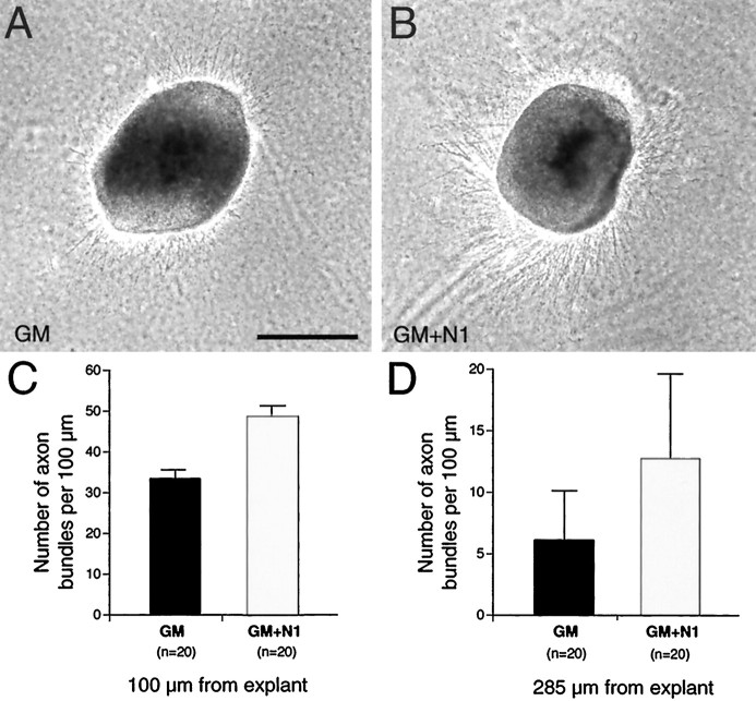
The thalamocortical axon (TCA) projection originates in dorsal thalamus, conveys sensory input to the neocortex, and has a critical role in cortical development. We show that the secreted axon guidance molecule netrin-1 acts in vitro as an attractant and growth promoter for dorsal thalamic axons and is required for the proper development of the TCA projection in vivo. As TCAs approach the hypothalamus, they turn laterally into the ventral telencephalon and extend toward the cortex through a population of netrin-1-expressing cells. DCC and neogenin, receptors implicated in mediating the attractant effects of netrin-1, are expressed in dorsal thalamus, whereas unc5h2 and unc5h3, netrin-1 receptors implicated in repulsion, are not. In vitro, dorsal thalamic axons show biased growth toward a source of netrin-1, which can be abolished by netrin-1-blocking antibodies. Netrin-1 also enhances overall axon outgrowth from explants of dorsal thalamus. The biased growth of dorsal thalamic axons toward the internal capsule zone of ventral telencephalic explants is attenuated, but not significantly, by netrin-1-blocking antibodies, suggesting that it releases another attractant activity for TCAs in addition to netrin-1. Analyses of netrin-1 -/- mice reveal that the TCA projection through the ventral telencephalon is disorganized, their pathway is abnormally restricted, and fewer dorsal thalamic axons reach cortex. These findings demonstrate that netrin-1 promotes the growth of TCAs through the ventral telencephalon and cooperates with other guidance cues to control their pathfinding from dorsal thalamus to cortex.


Figures

Fig. 1.
Fig. 1.
TCA pathway. Schematic of a coronal section of mouse brain showing the mature TCA projection and relevant brain structures. TCAs (black), originating in dorsal thalamus (dTh;light gray), project ventrally along the lateral aspect of ventral thalamus (vTh). As they approach the hypothalamus (Hy), TCAs make a sharp lateral turn and extend dorsolaterally through the striatum (St) toward neocortex (Ctx). TCAs are compactly bundled along the proximal portion of their path, but fan-out into numerous fascicles as they extend dorsolaterally through the striatum. As TCAs extend through the Ctx, their path becomes centered on the subplate layer (SP), and branches of TCAs (medium gray) extend into and innervate the cortical plate (CP). TCAs, together with the oppositely extending corticothalamic axons, form the internal capsule (ic), the major axon tract for cortical input and output projections.Dashed lines indicate the approximate borders between the ventral thalamus and hypothalamus, diencephalon and striatum, and striatum and neocortex.
Fig. 2.
Fig. 2.
Expression of netrin-1 and netrin receptors in relation to the TCA projection. Coronal sections of E14.5 (A, A′) and E13.5 (B–E) mouse brains showing netrin-1 (A, A′, B),DCC (C), neogenin(D), and unc5h3(E) expression detected with digoxigenin-labeled riboprobes. Dorsal is up. Lateral is to theright in A′ and B.A, At E14.5, netrin-1 expression is detected in the differentiating mantle zone of the ICZ, as well as in the overlying ventricular zone (small arrow).Netrin-1 is also expressed in the medial part of dorsal thalamus (dTh; small arrowhead), hippocampus (Hi; large arrow), and the hypothalamic (Hy) ventricular zone (large arrowhead). A′,Higher magnification of boxed area in A.This is the region used in coculture experiments in Figure 7 (see Materials and Methods). Netrin-1-expressing cells are present in the ICZ (arrows), intermingled with fascicles of TCAs in the internal capsule (ic). B,At E13.5, netrin-1 is expressed in the ICZ, in cells within (small arrow) and surrounding (large arrow) the internal capsule (ic). Expression is also detected in the overlying ventricular zone of the ganglionic eminence (arrowhead). C,DCC expression is detected in the ventricular zone (small arrow) and differentiating mantle zone (large arrow) of dorsal thalamus, and in cells at the dorsal thalamic-ventral thalamic border (arrowhead). The expression within the mantle zone corresponds to the location of the ventral posterior thalamic nucleus (VB).Asterisk indicates high expression in medial habenula.D,Neogenin is expressed in medial dorsal thalamus (arrow). E,Unc5h3 is expressed in the lateral dorsal part of ventral thalamus (arrow), but not in dorsal thalamus. Scale bars: A, 500 μm; A′,B, C, 250 μm (bar in Calso applies to D, E).
Fig. 3.
Fig. 3.
Netrin-1-dependent attraction of dorsal thalamic axons. Explants from the medial half of dorsal thalamus (dTh; see Materials and Methods; E13.5–E14.5 mouse) were cocultured for 1.5–2 d in collagen gels at a distance from floor plate (FP; E17–E18 rat) or aggregates of 293 cells transfected with netrin-1 (dTh-293Net) or the parental plasmid as a control (dTh-293).A, Dorsal thalamic axon outgrowth is biased toward FP.B, Axon outgrowth from dorsal thalamus is also biased toward FP when nonimmune sera (10 μg/ml) is added to the growth medium (GM). C, Axon outgrowth from dorsal thalamus is no longer biased toward FP when netrin-1 blocking antibodies (10 μg/ml) are added to the GM. D, E, Quantification of dorsal thalamic axon responses.N (number of cocultures) values are inparentheses. D, When dorsal thalamus is cultured alone (dTh alone), the distribution of axon outgrowth is significantly different from dorsal thalamus cocultured with FP (GM; p < 0.0002, χ2 test). When dorsal thalamus is cocultured with FP in the presence of netrin-1 blocking antibodies (anti-N1), the distribution of axon outgrowth is significantly different from dorsal thalamus cultured in GM, or GM with added nonimmune sera (control IgG; p < 0.004 and 0.02, respectively, χ2 test). Dorsal thalamic axon outgrowth in cocultures with control IgG was not significantly different from that with GM alone (p = 0.85, χ2test). E, Although there is a trend for dorsal thalamic axon outgrowth to be biased toward netrin-1-expressing cells (dTh-293Net) compared to control 293 cells (dTh-293), these differences are not significant (p = 0.5, χ2 test). Scale bar, 250 μm.
Fig. 4.
Fig. 4.
The path of TCAs is abnormally restricted in E15 netrin-1 −/− mice. Coronal sections of E15 mouse brains from wild-type (A–C) andnetrin-1 −/− (D–F) littermates with DiI implanted into dorsal thalamus, showing DiI-labeled TCAs (B, E), bisbenzimide counterstain of the same sections (A, D), and computer-generated overlays of the DiI and bisbenzimide images (C, F). In wild-type mice, TCAs are broadly and evenly distributed in the internal capsule (ic). In netrin-1 −/− mice, the TCA pathway is restricted dorsomedially, and fascicles of TCAs appear disorganized; the ventrolateral portion of the pathway is devoid of TCA fascicles (E, F, arrows). The magnitude of the TCA projection is also reduced in the netrin-1 −/− mice compared to their wild-type littermates. In both wild-type andnetrin-1 −/− littermates, TCAs have entered the intermediate zone/subplate underlying the neocortex (Ctx; B, E, arrowheads).Asterisks in A and Dindicate the rostral portion of the thalamus. Hi, Hippocampus; St, striatum. Scale bar, 300 μm. Dorsal is to the top, and lateral is to theright.
Fig. 5.
Fig. 5.
The internal capsule is abnormally narrow, and fascicles of TCAs are disorganized in P0 netrin-1−/− mice. Coronal sections of P0 mouse brains from wild-type (A–C) and netrin-1 −/− (D–F) littermates with DiI implanted into dorsal thalamus (dTh), showing DiI-labeled processes (B, E), bisbenzimide counterstain (A, D), and computer-generated overlay of DiI and bisbenzimide images (C, F). In wild-type mice, the fascicles of TCAs (B, C, between arrows) are distributed throughout most of the striatum. In P0 netrin-1 −/− mice, the fascicles of TCAs (E, F, between arrows) are restricted to dorsomedial striatum. In both wild-type and netrin-1−/− littermates, TCAs have extended into neocortex (Ctx) and have begun to invade the cortical plate (B, E, asterisks). G, H, Coronal sections through the caudal part of the internal capsule (ic) of P0 wild-type (G) and netrin-1−/− (H) littermates immunolabeled with an L1 antibody and visualized with a peroxidase-conjugated secondary antibody. In wild-type mice, the L1-immunolabeled axon fascicles are relatively thin and extend roughly parallel to one another (G, arrow). In netrin-1 −/− mice, the L1-immunolabeled axon fascicles are thicker, less numerous, and abnormally organized (H, arrow). I,Quantification of the distribution of DiI-labeled fascicles of TCAs within the internal capsule of P0 wild-type and netrin-1−/− mice. For quantification, mice were paired according to the size and placement of DiI in the dorsal thalamus. The width of the internal capsule (i.e., the distribution of labeled fascicles of TCAs, measuredbetween arrows in B, E) within the striatum in netrin-1 −/− mice is significantly reduced, being ∼50% of that in paired wild-type littermates (I; n = 4 littermate pairs, p < 0.001 Student's ttest). cc, Corpus callosum; pb, Probst bundle; wt, wild type. Scale bars: A, 400 μm (also applies to B–F); G, 300 μm (also applies to H). Dorsal is to thetop, and lateral is to the right.
Fig. 6.
Fig. 6.
The TCA projection is variably reduced innetrin-1 −/− mice. Coronal sections of E17.5 (A, B′) and E18.5 (C–H) mouse brains from wild-type (A, A′, C, C′, E, E′, G) andnetrin-1 −/− (B, B′, D, D′, F, F′, H) littermates, showing DiI-labeled cell bodies (G, H), axons (A′–D′, E, F), and bisbenzimide counterstain of the same sections (A–D, E′, F′). DiI was implanted into the ventroposterior thalamic nucleus (VP) to anterogradely label VP axons (A′–D′, E, F), or into somatosensory cortex to retrogradely label VP neurons (G, H). I, Quantification of retrogradely labeled VP neurons. In wild-type animals at E17.5 (A′) and E18.5 (C′), TCAs pass through the internalcapsule (ic) to reach cortex, extend intracortically within the subplate (SP;A′, B′, arrowheads), and extend branches into the cortical plate (CP). In contrast, innetrin-1 −/− littermates at E17.5 (B′) and E18.5 (D′), the TCA projection is reduced (B′, D′, arrowheads). E, F, Higher magnification images of the TCA projections shown in C′and D′, respectively. In the wild-type mouse, a substantial population of anterogradely labeled VP axons is present in the subplate, and their branches densely invade the cortical plate (E). In the netrin-1 −/− littermate, fewer labeled VP axons reach the subplate, and fewer branches extend into the cortical plate (F).G, H, The number of VP neurons retrogradely labeled by DiI injected into somatosensory cortex in netrin-1 −/− mice (H) is considerably reduced compared to their matched wild-type littermates (G).Dashed white lines mark the lateral border of the VP nucleus. I, The number of retrogradely labeled VP neurons in netrin-1 −/− mice plotted as a percentage of the number in wild-type littermates paired for injection size and location. Case #1 corresponds to the pair shown inG and H. The mean value for the five cases is also plotted. LG, Lateral geniculate nucleus;MZ, marginal zone; tr, thalamic radiations. Scale bars: A–D′, 200 μm (also applies to A–C and A′–C′); E, F′, 100 μm; G, H, 100 μm. Dorsal is to thetop, and lateral is to the right.
Fig. 7.
Fig. 7.
The ICZ releases an attractant activity for dorsal thalamic axons that is distinct from netrin-1. Explants from the medial half of dorsal thalamus (dTh; E13.5–E14.5 mouse) were cocultured for 1.5–2 d in collagen gels at a distance from the location of the ICZ in explants of ventral telencephalon (E13.5–E14.5 mouse). Netrin-1 blocking antibodies (anti-N1; 10 μg/ml) or nonimmune sera (control IgG; 10 μg/ml) were added to the growth medium at the time of plating. The number of cocultures (n) is indicated in parentheses. When cultured in the presence of control IgG, dorsal thalamic axon outgrowth is biased toward the ICZ (A, B). Addition of netrin-1 blocking antibodies diminishes the percentage of dorsal thalamic explants showing biased outgrowth toward the ICZ (B,anti-N1); however, the distributions of dorsal thalamic axon outgrowth in the control IgG and anti-N1 cocultures are not significantly different (p = 0.7, χ2 test). Scale bar, 250 μm.
Fig. 8.
Fig. 8.
Netrin-1 enhances dorsal thalamic axon outgrowth. Explants from the medial half of dorsal thalamus (dTh) were cultured in collagen gels for 1.5–2 d either in growth medium (A, GM) or in growth medium supplemented with soluble recombinant netrin-1 (B, GM+N1; 400 ng/ml). C, D, Quantification of axon outgrowth at 100 μm (C) and 285 μm (D) from dorsal thalamic explants. The number of cultures analyzed is given in parentheses. A greater number of axons are present at both 100 μm (C) and 285 μm (D) in the cultures to which netrin-1 had been added. The differences in axon outgrowth between the two culture types are significantly different (p < 0.0001 at 100 μm;p < 0.01 at 285 μm, Student's ttest). Scale bar, 250 μm.

References

    1. Blakemore C, Molnar Z. Factors involved in the establishment of specific interconnections between thalamus and cerebral cortex. Cold Spring Harb Symp Quant Biol. 1990;55:491–504. - PubMed
    1. Bloch-Gallego E, Ezan F, Tessier-Lavigne M, Sotelo C. Floor plate and netrin-1 are involved in the migration and survival of inferior olivary neurons. J Neurosci. 1999;19:4407–4420. - PMC - PubMed
    1. Braisted JE, Tuttle R, O'Leary DDM. Thalamocortical axons are influenced by chemorepellent and chemoattractant activities localized to decision points along their path. Dev Biol. 1999;208:430–440. - PubMed
    1. Butler H, Juurlink BHJ. An atlas for staging mammalian and chick embryos. CRC; Boca Raton: 1987.
    1. Catalano SM, Shatz CJ. Activity-dependent cortical target selection by thalamic axons. Science. 1998;281:559–562. - PubMed

Publication types

MeSH terms