. 2000 Aug 1;97(16):8921-5.
doi: 10.1073/pnas.150116497.

The molecular basis of vancomycin resistance in clinically relevant Enterococci: crystal structure of D-alanyl-D-lactate ligase (VanA)

Affiliations

The molecular basis of vancomycin resistance in clinically relevant Enterococci: crystal structure of D-alanyl-D-lactate ligase (VanA)

D I Roper et al. Proc Natl Acad Sci U S A. .

Abstract

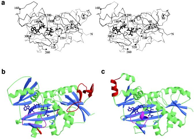
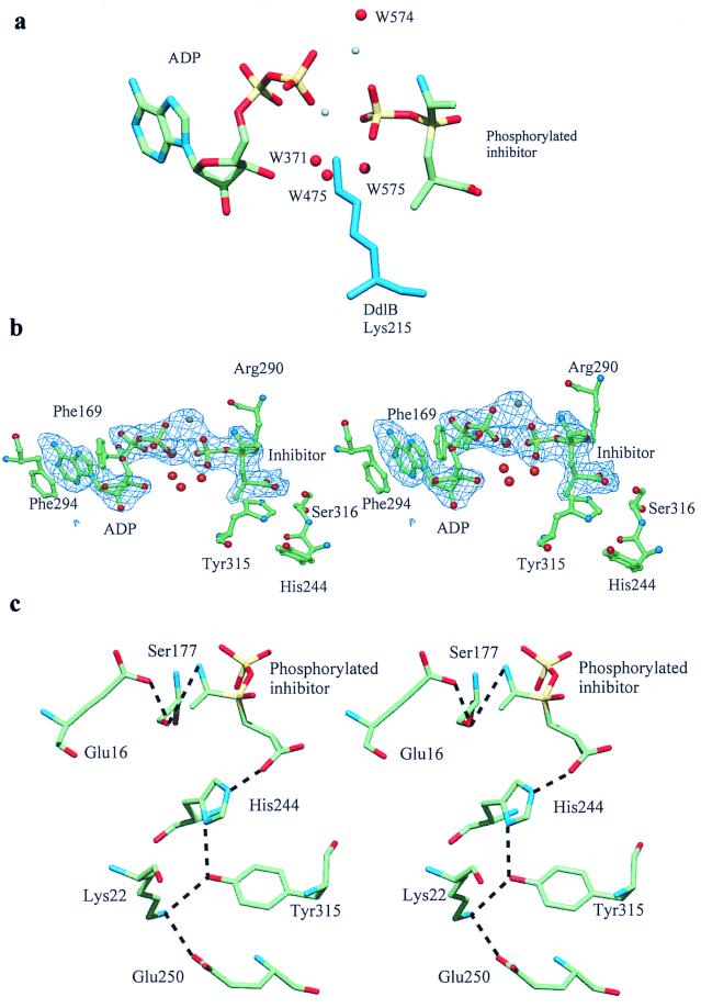
d-alanine-d-lactate ligase from Enterococcus faecium BM4147 is directly responsible for the biosynthesis of alternate cell-wall precursors in bacteria, which are resistant to the glycopeptide antibiotic vancomycin. The crystal structure has been determined with data extending to 2.5-A resolution. This structure shows that the active site has unexpected interactions and is distinct from previous models for d-alanyl-d-lactate ligase mechanistic studies. It appears that the preference of the enzyme for lactate as a ligand over d-alanine could be mediated by electrostatic effects and/or a hydrogen-bonding network, which principally involve His-244. The structure of d-alanyl-d-lactate ligase provides a revised interpretation of the molecular events that lead to vancomycin resistance.


Figures

Figure 1
Figure 1
Stereo C-α trace of VanA (a) with ADP and phosphophosphinate inhibitor showing overall structural features. Every twentieth residue is highlighted with a closed circle. N and C termini are marked N and C, respectively. In b and c, the structures of VanA and DdlB were overlaid in the program quanta and aligned by using close-residue optimization. Helices are displayed in green and β sheets in blue. ADP and phosphinophosphinate inhibitor are displayed in purple and magnesium ions in yellow. The ω-loops of both VanA and DdlB are displayed in magenta to highlight structural differences. Structural features corresponding to amino acid sequence found in one protein compared with the other are shown in red. In the structural alignment, a large additional loop occurs in VanA, between residues 44 and 90. The N terminus of each protein is shown on the right of each structure, immediately before the beginning of a β sheet found in both DdlB and VanA.
Figure 2
Figure 2
Schematic diagram of the active site of VanA from the structure showing the interaction of various water molecules in addition to amino acid side chains. The protein backbone of the residues in the region of His-244 and Tyr-315 is shown as a solid black line. Hydrogen bond distances between Tyr-315, His-244, and the second subsite carboxyl oxygen of the transition state inhibitor are 2.77 Å and 2.74 Å, respectively. Two phenylalanine residues (F169 and F294) form stacking interactions with the adenine nucleotide rings. Several water molecules in the active site form interactions with the phosphosphate and magnesium atoms as well as amino acid side chains. There is an additional hydrogen bond between water 475 and water 371, which is not shown for clarity.
Figure 3
Figure 3
(a) Comparison of active site waters in E. facieum VanA, which replace Lys-215 in E. coli DdlB. Magnesium atoms are shown in gray with water molecules in red. The structures of VanA and DdlB were superimposed and the relative positions of waters in VanA and Lys-215 in DdlB compared. Water 475 in VanA takes an equivalent position to the side-chain nitrogen of Lys-215 in DdlB. Hydrogen-bonding distances between adjacent water molecules, magnesium atoms, and phosphate atoms are not shown for clarity. Other water molecules, notably W371, W569, and W574, coordinate with magnesium ions in the active site of VanA. (b) A stereo representation and 2 FoFc map showing the active-site residues in contact with the phosphinophosphinate transition state intermediate. The map is contoured at 1σ by using the final 2.5-Å resolution map. Residues in the immediate vicinity of the transition state intermediate are marked. The two magnesium ions that coordinate with the phosphate ion of the intermediate and the β-phosphate of ADP are displayed in gray, and water molecules in this vicinity are displayed in red. Water 475 in VanA takes an equivalent position to the side-chain nitrogen of Lys-215 in DdlB. Other water molecules, notably W371 and W574, coordinate with magnesium ions in the active site of VanA. (c) Stereo diagram of the hydrogen bonding interacts with and in the vicinity of the phosphorylated phosphinate inhibitor in the active site. The Glu-250, Lys-22, Tyr-4315, and His-244 hydrogen-bonding network is shown, making a 2.7-Å hydrogen bond with the carboxylate oxygen of the inhibitor. This carboxylate also hydrogen bonds to a conserved serine (316 in VanA), which is not shown for clarity. The position of His-244 in our structure is such that it cannot make hydrogen-bonding interactions with the Glu-16 and Ser-177, which are structurally conserved in comparison to DdlB. The later two amino acids form important interactions that anchor d-Ala in the first subsite, an analogous situation to that found in DdlB.

References

    1. Novak R, Henriques B, Charpentier E, Normark S, Tuomanen E. Nature (London) 1999;399:580–593.
    1. Walsh C T, Fisher S L, Park I-S, Prahalad M, Wu Z. Chem Biol. 1996;3:21–28. - PubMed
    1. Bugg T D H, Wright G, Dutka-Malen S, Arthur M, Courvalin P, Walsh C T. Biochemistry. 1991;30:10408–10415. - PubMed
    1. Park I-S, Lin C-H, Walsh C T. Biochemistry. 1996;35:10464–10471. - PubMed
    1. Reynolds P E, Depardieu F, Dutka-Malen S, Arthur M, Courvalin P. Mol Microbiol. 1994;13:1065–1070. - PubMed

Publication types

MeSH terms

Associated data

LinkOut - more resources