. 2000 Aug 1;97(16):9305-10.
doi: 10.1073/pnas.160128797.

Specific binding sites for alcohols and anesthetics on ligand-gated ion channels

Affiliations

Specific binding sites for alcohols and anesthetics on ligand-gated ion channels

M P Mascia et al. Proc Natl Acad Sci U S A. .

Abstract

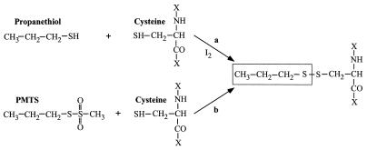
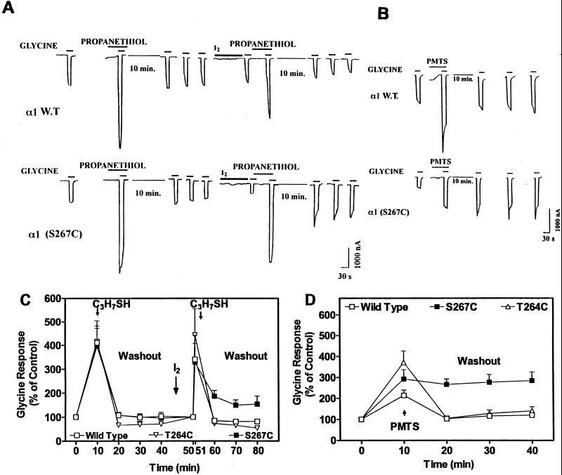
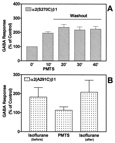
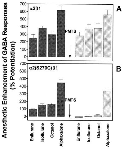
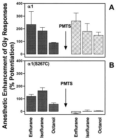
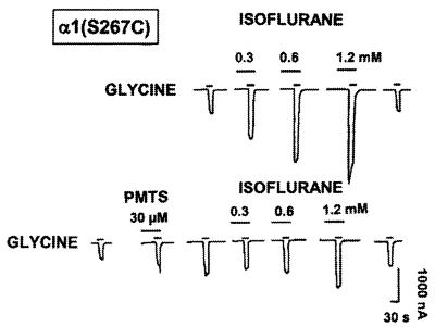
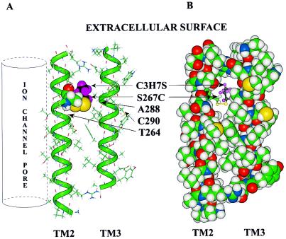
Ligand-gated ion channels are a target for inhaled anesthetics and alcohols in the central nervous system. The inhibitory strychnine-sensitive glycine and gamma-aminobutyric acid type A receptors are positively modulated by anesthetics and alcohols, and site-directed mutagenesis techniques have identified amino acid residues important for the action of volatile anesthetics and alcohols in these receptors. A key question is whether these amino acids are part of an alcohol/anesthetic-binding site. In the present study, we used an alkanethiol anesthetic to covalently label its binding site by mutating selected amino acids to cysteine. We demonstrated that the anesthetic propanethiol, or alternatively, propyl methanethiosulfonate, covalently binds to cysteine residues introduced into a specific second transmembrane site in glycine receptor and gamma-aminobutyric acid type A receptor subunits and irreversibly enhances receptor function. Moreover, upon permanent occupation of the site by propyl disulfide, the usual ability of octanol, enflurane, and isoflurane to potentiate the function of the ion channels was lost. This approach provides strong evidence that the actions of anesthetics in these receptors are due to binding at a single site.


Figures

Figure 1
Figure 1
Reaction of propanethiol and PMTS with the sulfhydryl group of cysteine. Note that the side chain structure attached to the cysteine residue after the reaction is identical for both reagents.
Figure 2
Figure 2
Effects of propanethiol or PMTS on α1 wild-type, α1(S267C), and α1(T264C) GlyR expressed in Xenopus oocytes. (A and B) Sample tracings of currents induced by application of an EC5 concentration of glycine alone or in the presence of propanethiol (4 mM) and iodine (0.5 mM) or PMTS (30 μM) in individual voltage-clamped oocytes. The time course of the washout of propanethiol (either before and after iodine application) or PMTS was measured every 10 min for a total of 30 min. (C) Propanethiol (4 mM) reversibly enhanced the glycine response in α1 wild-type (□), α1 (T264C) (▿), or α1 (S267C) (■) receptors before application with iodine (0.5 mM). After sequential application of iodine and propanethiol, the enhancement of the glycine response by propanethiol is reversible in wild-type and T264C but not in S267C receptors. Values are expressed as percentage of control. Control values determined at 0 time were set at 100% and were used to calculate data for 10–40 min; values determined at 50 min (immediately after application of I2) were set at 100% and used to calculate data for 51–80 min. Two-way ANOVA indicated significant differences between wild-type and S267C receptors in the GlyR responses after the application of iodine and propanethiol (P < 0.05). (D) PMTS (30 μM) enhanced the glycine response in all of the receptors and this was reversed to control level by washout in wild-type (□) and in T264C (▵) GlyR. In S267C receptors (■), the enhancement of the glycine response by PMTS was not reversible. Propanethiol or PMTS were perfused for 60 s before being coapplied with glycine for 30 s. The time course of the washout from propanethiol or PMTS was measured every 10 min for a total of 30 min. Two-way ANOVA indicated significant differences between wild-type and S267C receptors in the glycine responses after the application of PMTS (P < 0.001). For some points, error bars are smaller than symbols. Data are the mean ± SEM of five to nine oocytes.
Figure 3
Figure 3
Effects of PMTS on α2 (S270C)β1 and α2 (S291C)β1 GABAA receptors expressed in Xenopus oocytes. (A) In α2 (S270C)β1 GABAA receptors, PMTS (50 μM) irreversibly enhanced the GABA response. Washout from PMTS was measured every 10 min for a total of 30 min. (B) In α2 (S291C)β1 GABAA receptors, PMTS did not affect the GABA response. Isoflurane (0.6 mM) potentiated α2 (S291C)β1 GABA receptor function equally before and after application of PMTS. PMTS or isoflurane were perfused for 60 s before being coapplied with GABA for 30 s. Data are the mean ± SEM of five to six oocytes.
Figure 4
Figure 4
Effects of anesthetics on α2β1 or α2(S270C)β1 GABAA receptors, expressed in Xenopus oocytes, before and after perfusion with PMTS. (A) In wild-type α2β1 receptors, enflurane (1.2 mM), isoflurane (0.6 mM), octanol (0.1 mM), or alphaxalone (1 μM) enhanced GABA responses before and after application of PMTS (50 μM). (B) In mutant α2(S270C)β1 receptors, application of PMTS abolished the effects of enflurane, isoflurane, or octanol but did not affect the enhancement by alphaxalone. PMTS was perfused for 60 s before being coapplied with GABA for 30 s. Anesthetics or octanol were tested again after 15 min of washout from PMTS. Data are expressed as a mean ± SEM of five to 12 oocytes.
Figure 5
Figure 5
Effects of anesthetics on α1 or α1(S267C) GlyR, expressed in Xenopus oocytes, before and after perfusion with PMTS. (A) In α1 receptors, enflurane (1.2 mM), isoflurane (0.6 mM), or octanol (0.1 mM) enhanced glycine responses before and after application of PMTS (30 μM). (B) In mutant α1(S267C) receptors, application of PMTS abolished the effects of enflurane, isoflurane, or octanol. PMTS was perfused for 60 s before being coapplied with glycine for 30 s. Anesthetics or octanol were tested again after 15 min of washout from PMTS. Data are expressed as a mean ± SEM of three to four oocytes.
Figure 6
Figure 6
Effects of isoflurane on α1(S267C) mutant GlyR expressed in Xenopus oocyte. Isoflurane (0.3–1.2 mM) potentiates in a concentration-dependent manner the function of α1(S267C) mutant receptor before application of PMTS (30 μM). Application of PMTS abolished or reduced the potentiation of the GlyR by isoflurane. Sample tracings of currents were induced by application of an EC10 concentration of glycine alone or glycine in the presence of isoflurane in an individual voltage-clamped oocyte. Tracing is from a single oocyte, and similar results were obtained from two other oocytes.
Figure 7
Figure 7
Molecular model of TM2 and TM3 of the GlyR α1 subunit. TM2 is oriented so that the aqueous pore of the ion channel would be on the far left (vertical cylinder). This orientation places residue S267C on the interior of the subunit. The TM3 helix was aligned anti-parallel to TM2 so that S267 and A288 would be on the same horizontal plane. (A) The peptides are rendered as a stick structure, the backbones of the α helices are overlaid with a green tube, and both S267C and propanethiol are rendered with a space-filling (van der Waals) surface. The colors of atoms are: carbon, green; hydrogen, gray; oxygen, red; nitrogen, blue; and the propyl group of propanethiol is highlighted with black carbons and violet hydrogens. Those cysteine residues that did not form covalent bonds with PMTS or that formed them without any effect on potentiation of the effect of glycine are indicated (T264C, A288C, and C290). (B) The same molecular model of TM2 and TM3 is shown except that all residues in the two helices are rendered with a space-filling surface and the propanethiol is rendered as a ball-and-stick molecule. It can be seen that the propyl group occupies the cavity and may prevent the additional occupation of the cavity by octanol, isoflurane, or enflurane.

References

    1. Kennedy S K, Longnecker D E. In: The Pharmacological Basis of Therapeutics. Moliniff P B, Ruddon R W, editors. New York: McGraw–Hill; 1996. pp. 304–305.
    1. Meyer H H. Arch Exp Pathol Pharmakol. 1899;42:109–118.
    1. Overton E. Studien uber die Narkose, Zugleich ein Beitrag zur Allgemeinen Pharmakologie. Jena, Germany: Gustav Fischer; 1901. pp. 1–195.
    1. Cantor R S. Biochemistry. 1997;36:2339–2344. - PubMed
    1. Forman S A, Miller K W, Yellen G. Mol Pharmacol. 1995;48:574–581. - PubMed

Publication types

LinkOut - more resources