. 2000 Aug 1;97(16):9144-9.
doi: 10.1073/pnas.160258197.

Engineering blood meal-activated systemic immunity in the yellow fever mosquito, Aedes aegypti

Affiliations

Engineering blood meal-activated systemic immunity in the yellow fever mosquito, Aedes aegypti

V Kokoza et al. Proc Natl Acad Sci U S A. .

Abstract

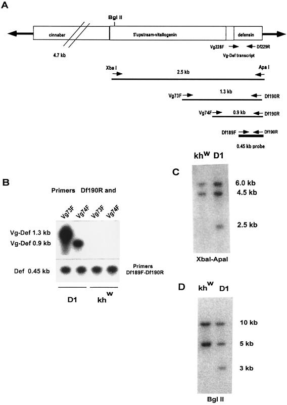
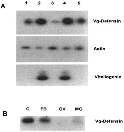
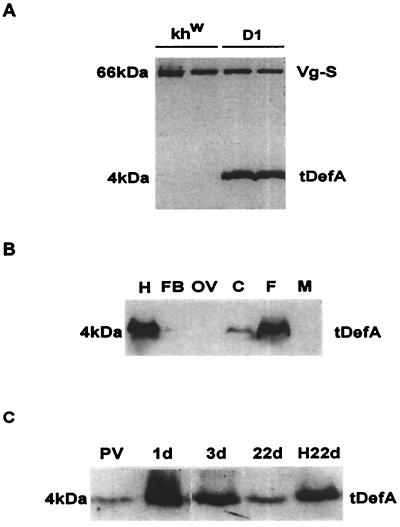
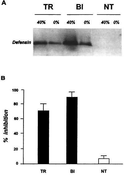
Progress in molecular genetics makes possible the development of alternative disease control strategies that target the competence of mosquitoes to transmit pathogens. We tested the regulatory region of the vitellogenin (Vg) gene of Aedes aegypti for its ability to express potential antipathogen factors in transgenic mosquitoes. Hermes-mediated transformation was used to integrate a 2.1-kb Vg-promoter fragment driving the expression of the Defensin A (DefA) coding region, one of the major insect immune factors. PCR amplification of genomic DNA and Southern blot analyses, carried out through the ninth generation, showed that the Vg-DefA transgene insertion was stable. The Vg-DefA transgene was strongly activated in the fat body by a blood meal. The mRNA levels reached a maximum at 24-h postblood meal, corresponding to the peak expression time of the endogenous Vg gene. High levels of transgenic defensin were accumulated in the hemolymph of bloodfed female mosquitoes, persisting for 20-22 days after a single blood feeding. Purified transgenic defensin showed antibacterial activity comparable to that of defensin isolated from bacterially challenged control mosquitoes. Thus, we have been able to engineer the genetically stable transgenic mosquito with an element of systemic immunity, which is activated through the blood meal-triggered cascade rather than by infection. This work represents a significant step toward the development of molecular genetic approaches to the control of vector competence in pathogen transmission.


Figures

Figure 1
Figure 1
Stable integration of the Vg-Defensin transgene into the Aedes aegypti genome. (A) Schematic diagram of the transformation vector construct, pH[cn][Vg-DefA], used in this study to transform mosquitoes. The Hermes inverted terminal repeats are represented as solid arrows flanking the 4.7-kb genomic DNA fragment of the D. melanogaster cn+ marker gene and the 2.5-kb Vg-DefA fusion gene as the promoter–reporter part of the construct. The arrows and numbers below the diagram show the relative extents and the expected sizes of the fragments generated by XbaI–ApaI restriction digest and by gene amplification using different pairs of primers specific to the Vg-DefA fusion sequence. The 0.45-kb DefA DNA sequence used as a probe in these experiments is shown as a black box below the map. (B) Gene amplification analysis of genomic DNA isolated from G4 mosquito progeny of the D1 transgenic line and the control khw strain. To confirm the specificity of amplification for the Vg-DefA transgene, the gene amplification products were analyzed by Southern blot hybridization using the DefA DNA as a probe. By using the Vg-DefA primer combinations, Vg73F-Df190R and Vg74F-Df190R, the transgenic mosquito DNA showed specific amplification of fragments of the expected sizes, 1.3-kb and 0.9-kb, respectively. No amplification was observed in the genomic DNA from the control strain (Upper). As a positive control for loading and for the integrity of isolated genomic DNA, DefA-specific primer pairs, Df189F-Df190R, were used, and these produced a band of the expected size, 0.45-kb, in all samples (Lower). (C) Southern blot analysis of genomic DNA prepared from G4 progeny of transformed mosquitoes of the D1 line and the host strain (khw), digested with XbaI–ApaI and hybridized with the DefA DNA probe. A diagnostic hybridization signal of the expected size, 2.5 kb, is associated with the insertion of the Vg-DefA transgene into the mosquito genome and is evident in DNA isolated from transformed mosquitoes, but not in the host strain. The two higher molecular weight signals of 6 kb and 4.5 kb, seen in DNA digests of both the transgenic and the host strains, represented two copies of endogenous DefA genes. (D) Southern blot analysis of genomic DNA isolated from G9 progeny of the D1 transgenic line and the host khw strain, digested with BglII and hybridized with DefA DNA probe. The unique hybridization signal of 3.0-kb associated with Vg-DefA insertion is seen only in the D1 transgenic line.
Figure 2
Figure 2
RT-PCR analyses of the Vg-DefA transgene expression. (A) Blood meal activation of the Vg-DefA transgene in mRNA samples isolated from transgenic males (1), blood-fed females (2), previtellogenic (3) and vitellogenic females 24 (4), and 72 hPBM (5). The same RNA samples also were analyzed using actin-specific primers as a control for RNA integrity and loading (Middle), and vitellogenin-specific primers, as a control showing the expression of the endogenous Vg gene after blood meal activation (Bottom). (B) Tissue-specific expression of the Vg-DefA transgene in vitellogenic females. Specific amplification was observed only in RNA samples isolated from the fat body (FB) and carcass (C), containing the fat body of the thorax and head. No amplification was found in the ovary (OV) or midgut (MG).
Figure 3
Figure 3
Immunoblot analyses of tDefA peptide expression in transgenic mosquitoes after blood meal activation. (A) Protein extracts from individual 24 hPBM females of the host khw strain and the D1 transgenic line were tested by using polyclonal antibody to Ae. aegypti DefA and mAb to Ae. aegypti vitellogenin small subunit (27). Expression of the 4-kDa DefA peptide was observed in transgenic mosquitoes but not in the khw host strain. The expression of 66-kDa Vg-small subunit (Vg-S), used as a positive control for blood meal activation, was present at the same level in both the transgenic line and the host strain. (B) Immunoblot analysis of tissue- and sex-specific expression of tDefA peptide in transgenic mosquitoes. Protein extracts from hemolymph (H), fat bodies (FB), ovary (OV), carcass (C), 24 hPBM whole mosquito female, (F), and mosquito male (M) were analyzed. (C) Time course of tDefA protein accumulation in transgenic mosquitoes during the vitellogenic cycle after a single blood feeding. Previtellogenic female (PV), vitellogenic females 1 (1d), 3 (3d), and 22 (22d) days PBM and the hemolymph collected from 22-day-old female mosquitoes (H22d) were tested using DefA-specific antibodies. One mosquito-equivalent was loaded in each lane, except for the hemolymph sample from 22-day-old females, in which a four-mosquito equivalent was used.
Figure 4
Figure 4
Analysis of antibacterial activity of tDefA peptide isolated from transgenic mosquitoes. (A) Immunoblot analysis of peptide fractions used to assay the antibacterial activity. Protein samples obtained from transgenic 24 hPBM females of D1 line (TR), bacterially induced nontransgenic females of the khw host strain (BI), and nontransgenic 24 hPBM females of the khw host strain (NT) were isolated by using acid extraction and chromatography purification on Sep-Pak C18 cartridges and analyzed by immunoblotting. The analyzed samples were prepared from the 0% acetonitrile fraction in acidified water (0%) as a loading control, and the 40% acetonitrile fraction (40%), enriched for DefA peptide. (B) Aliquots from 40% fractions containing the major portion of defensin, isolated from transgenic (TR) and bacteria-induced (BI) females, were analyzed by the liquid growth inhibition assay using the Gram-positive bacteria, Micrococcus luteus. An equal aliquot of the 40% acetonitrile fraction isolated from nontransgenic 24 hPBM females of the host strain (NT) was used as a negative control. Data represent the mean of three independent experiments.

Comment in

References

    1. Butler D. Nature (London) 1997;386:535–536. - PubMed
    1. Bruno J M, Feachem R, Godal T, Nchinda T, Ogilvie B, Mons B, Mshana R, Radda G, Samba E, Schwartz M, et al. Nature (London) 1997;386:541. - PubMed
    1. Beier J C. Annu Rev Entomol. 1998;43:519–543. - PubMed
    1. Collins F H, James A A. Sci Med. 1996;3:52–61.
    1. O'Brochta D A, Atkinson P W. Insect Biochem Mol Biol. 1996;26:739–753. - PubMed

Publication types

LinkOut - more resources