. 2000 Aug 1;97(16):9015-20.
doi: 10.1073/pnas.160266597.

Protein-interaction modules that organize nuclear function: FF domains of CA150 bind the phosphoCTD of RNA polymerase II

Affiliations

Protein-interaction modules that organize nuclear function: FF domains of CA150 bind the phosphoCTD of RNA polymerase II

S M Carty et al. Proc Natl Acad Sci U S A. .

Abstract

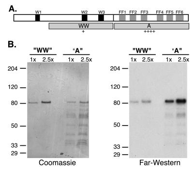
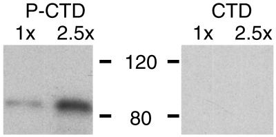
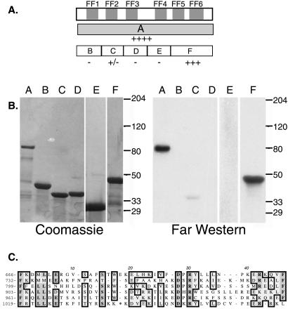
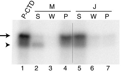
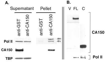
An approach for purifying nuclear proteins that bind directly to the hyperphosphorylated C-terminal repeat domain (CTD) of RNA polymerase II was developed and used to identify one human phosphoCTD-associating protein as CA150. CA150 is a nuclear factor implicated in transcription elongation. Because the hyperphosphorylated CTD is a feature of actively transcribing RNA polymerase II (Pol II), phosphoCTD (PCTD) binding places CA150 in a location appropriate for performing a role in transcription elongation-related events. Several recombinant segments of CA150 bound the PCTD. Predominant binding is mediated by the portion of CA150 containing six FF domains, compact modules of previously unknown function. In fact, small recombinant proteins containing the fifth FF domain bound the PCTD. PCTD binding is the first specific function assigned to an FF domain. As FF domains are found in a variety of nuclear proteins, it is likely that some of these proteins are also PCTD-associating proteins. Thus FF domains appear to be compact protein-interaction modules that, like WW domains, can be evolutionarily shuffled to organize nuclear function.


Figures

Figure 1
Figure 1
Identification of CA150 as a PCTD associating protein. (A) To identify PCTD-binding proteins from HeLa cells, we purified HeLa nuclear proteins and assessed their PCTD-binding abilities by direct binding in a Far Western analysis. The flow chart indicates the steps in the purification of a PCAP of molecular weight 150,000. (B) The Coomassie-stained proteins and PCTD interaction of 0.2 M NaCl elution of Q column demonstrate that the protein of molecular weight 150,000 binds the PCTD. Mass spectrometric analysis revealed that this protein is CA150.
Figure 2
Figure 2
Domains and PCTD binding of CA150. (A) Upper open box represents the 1,098-aa CA150 protein, with WW domains shaded black and FF motifs, numbered sequentially, shaded gray. Gray boxes in second row labeled WW and A represent segments of protein expressed in bacteria and tested for PCTD binding in B. Construct names and amino acid numbers: WW (); A (631–1098). Relative binding affinities to the PCTD, as judged by eye from Far Western analyses, are indicated below each construct. (B) GST fusion proteins carrying segments WW and A were run in two different amounts (1× = ≈100 ng for WW and ≈50 ng for A) on duplicate SDS gels that were either stained (Coomassie) or blotted to nitrocellulose and probed with 32P-labeled PCTD (Far Western). Prestained marker positions are indicated at left (apparent Mr). The amino terminal-most segment, containing WW1, was not successfully expressed in bacteria.
Figure 3
Figure 3
The FF region binding is specific for the phosphorylated CTD. Construct A of Fig. 2 was run in duplicate in two different amounts on an SDS gel, and the proteins were electroblotted to nitrocellulose strips. One strip was reacted with hyperphosphorylated βgal-yCTD fusion protein (PCTD) and the other with nonphosphorylated βgal-yCTD fusion protein (CTD). Bound fusion protein was visualized by using anti-βgal antibodies and chemiluminescence.
Figure 4
Figure 4
CA150 fragments bind PCTD by Far Western. (A) The upper box represents the C-terminal region of CA150 with the FF domains shaded gray. The gray box on the next line represents construct A. The open boxes on the third line represent constructs B–F. Construct names and amino acid numbers: A (631–1098); B (); C (); D (); E (); F (952–1098). Relative binding affinities to PCTD are indicated below each construct. (B) Constructs A–F were separated on duplicate 4–15% SDS gels. One gel was stained with Coomassie, whereas the duplicate was transferred to Nitrocellulose and probed with 32PCTD (Far Western). (C) Alignment of the FF motifs of CA150. The starting amino acid of each motif is indicated at left. Homologous amino acids are boxed and * in FF6 = LIQESDQHL.
Figure 5
Figure 5
FF5 (fragment M, amino acids 952-1027; see Table 1) binds the PCTD in solution. We bound the M (FF5) and the J (control, amino acids 1005–1027; Table 1) GST-fusion proteins to glutathione beads and then tested the ability of the bead-bound proteins to pull down 32PCTD (Methods). The 32PCTD in each sample was visualized by PhosphorImager. Lane 1 is the starting 32PCTD solution. Lanes 2–4 are the supernatant, wash, and pellet for sample M. Lanes 5–7 are the supernatant, wash, and pellet for sample J. Hyperphosphorylated CTD is indicated by the arrow and hypophosphorylated CTD is indicated by the arrowhead.
Figure 6
Figure 6
RNA polymerase II coimmunoprecipitates with CA150 from HeLa nuclear extracts. (A) CA150 was immunoprecipitated by using CA150 antibodies. GST antibodies were used as a negative control. Equal amounts of supernatants and pellets from the immunoprecipitations were separated by SDS/PAGE and subsequently analyzyed by immunoblotting with specific antibodies against RNA polymerase II (Pol II), CA150, and the TATA-binding protein (TBP). Both the hyperphosphorylated IIO form and the hypophosphorylated IIA form of Pol II largest subunit were detected in the anti-CA150 pellets. (B) The C-terminal half of CA150, containing the six FF repeats, is sufficient for binding to Pol II in vivo. Full length CA150 (FL) or the C-terminal half of CA150 (C) were expressed in 293T cells and immunoprecipitated via an amino-terminal T7 epitope tag. As a negative control, immunoprecipitates from 293T cells transfected with the expression vector alone (V) were also analyzed. The immunoprecipitates were separated by SDS/PAGE and immunoblotted with T7 tag antibodies, to detect CA150 proteins and with Pol II antibodies to detect subunits IIa/IIo (not resolved on this gel).

References

    1. Corden J L. Trends Biochem Sci. 1990;15:383–387. - PubMed
    1. Lu H, Flores O, Weinmann R, Reinberg D. Proc Natl Acad Sci USA. 1991;88:10004–10008. - PMC - PubMed
    1. Chesnut J D, Stephens J H, Dahmus M E. J Biol Chem. 1992;267:10500–10506. - PubMed
    1. Hampsey M, Reinberg D. Curr Opin Genet Dev. 1999;9:132–139. - PubMed
    1. Myer V E, Young R A. J Biol Chem. 1998;273:27757–27760. - PubMed

Publication types

LinkOut - more resources