Review
. 2000 Jun;53(6):409-18.
doi: 10.1136/jcp.53.6.409.

Problematic pigmented lesions: approach to diagnosis

Affiliations
Review

Problematic pigmented lesions: approach to diagnosis

S L Edwards et al. J Clin Pathol. 2000 Jun.

Abstract

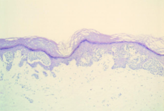
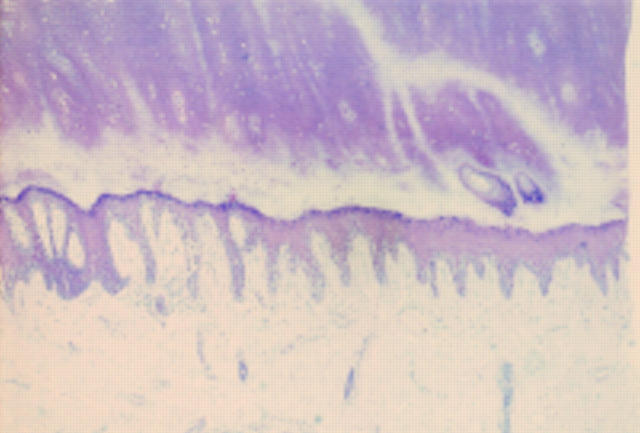
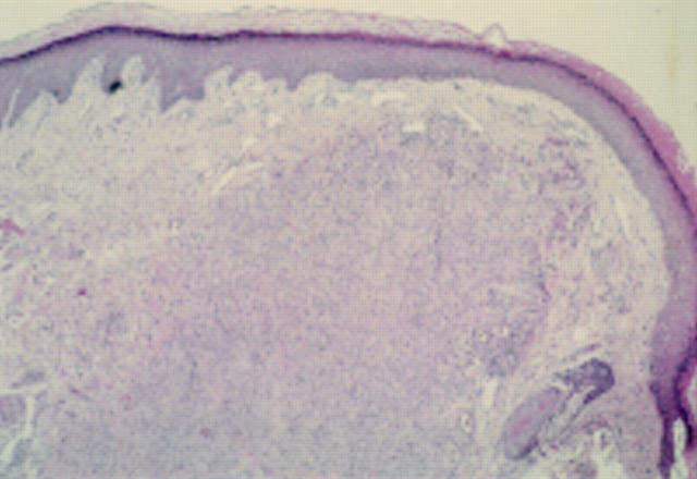
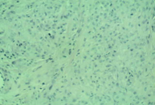
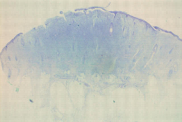
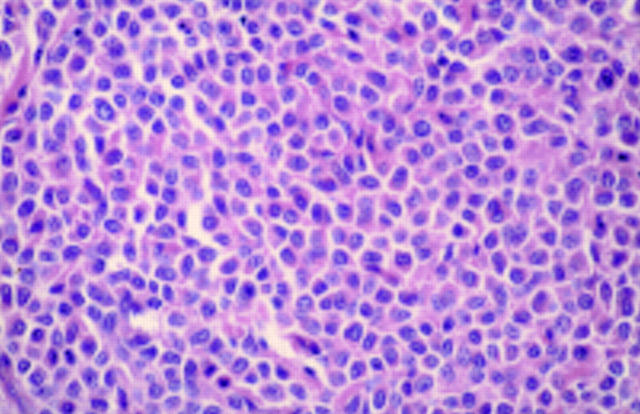
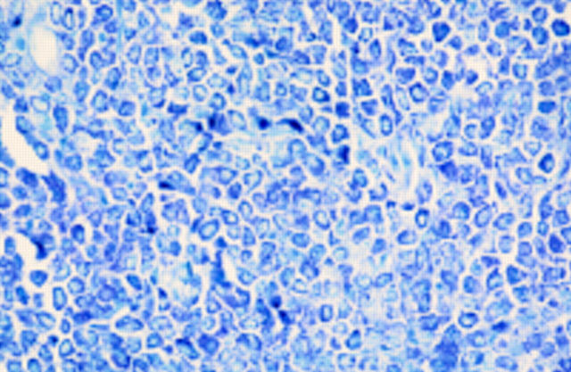
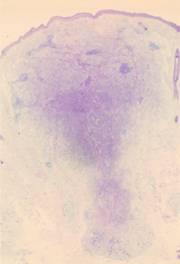
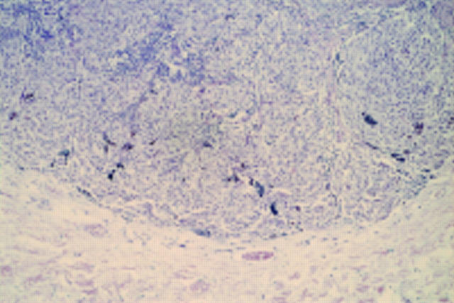
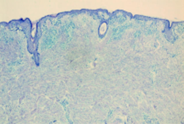
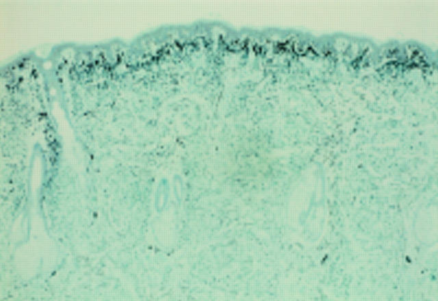
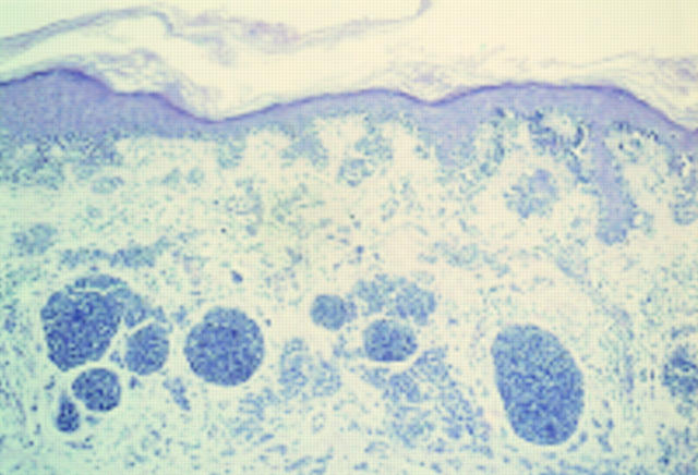
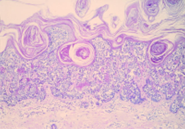
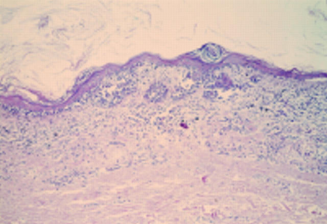
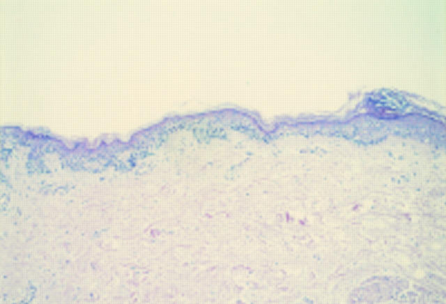
A number of pigmented lesions are difficult to classify and raise the possibility of a melanoma diagnosis. Care should be exercised to exclude non-melanocytic lesions, and benign melanocytic entities, both of which can mimic melanoma histologically. In addition, the possibility of the lesion being a melanoma variant or epidermotropic metastasis should be considered. There will still be some cases that are difficult to resolve. These usually fall into one of three categories: atypical junctional melanocytic lesion versus early melanoma; naevus versus naevoid melanoma; and atypical Spitz, cellular blue, and deep penetrating naevi versus thick melanoma. These will pose problems even for experts. The atypical Spitz lesions are perhaps the most important category because they tend to be from younger individuals, the differential diagnosis is thick melanoma, and there is no single discriminating histological feature.


Figures

None
Figure 1 Acral naevus. At low power there is pronounced junctional activity. No psoriasiform hyperplasia is seen.
None
Figure 2 Acral lentiginous melanoma in situ. At low power there is psoriasiform hyperplasia of the epidermis with a subtle lentiginous proliferation of melanocytes along the basal layer.
None
Figure 3 Epithelioid cell histiocytoma. A cellular dermal nodule with a Grenz zone. The lesion extends into deeper tissues.
None
Figure 4 Epithelioid cell histiocytoma. Cells are epithelioid and there is no apparent maturation. A mitotic figure is present. A recent case at the Scottish melanoma group (with permission from Dr KM McLaren, Edinburgh). The differential diagnosis is atypical Spitz, atypical cellular blue naevus, and metastatic melanoma. The lesion was S100 protein negative.
None
Figure 5 Mastocytoma. A cellular dermal lesion with overlying ulceration and extension of the cells into the fat.
None
Figure 6 Mastocytoma. Constituent cells of fig 5. The cells have a moderate eosinophilic cytoplasm. There is mild to moderate nuclear atypia.
None
Figure 7 Mastocytoma. Toluidine blue staining of cells. Cells were S100 protein and Melan-A negative.
None
Figure 8 Deep penetrating naevus. Symmetrical dermal lesion extending deeply into the fat. A case from the melanoma slide club (with permission from Dr H Rigby, Bristol). The differential diagnosis is atypical blue naevus and melanoma.
None
Figure 9 Deep penetrating naevus. The base of the lesion, which is rounded. The cells are growing in a fascicular pattern and there are mild cytological abnormalities. Deep or excessive mitotic activity would raise concerns about the lesion being a melanoma.
None
Figure 10 Desmoplastic melanoma. A junctional component is obvious at low power, but the dermis appears to be uninvolved.
None
Figure 11 Desmoplastic melanoma. The S100 protein shows the extent of the lesion within the dermis.
None
Figure 12 Small cell (non-Merkel) melanoma. There is an atypical junctional component and within the dermis there are nests composed of hyperchromatic cells with scanty cytoplasm and mild cytological atypia. There is a proliferation of small blood vessels between the nests and a patchy lymphocytic infiltrate. The patient was in their sixth decade.
None
Figure 13 Verrucous small cell melanoma. At scanning power there is a warty looking, rather bland melanocytic lesion, composed of small naevoid like cells.
None
Figure 14 Verrucous small cell melanoma. An area of regression within the lesion.
None
Figure 15 Verrucous small cell melanoma. The lateral growth spread of the lesion. Taken together with the age of the patient, the regression favoured a malignant diagnosis.

References

    1. Arch Dermatol. 1975 Dec;111(12):1588-90 - PubMed
    1. Am J Surg Pathol. 1989 Jan;13(1):39-44 - PubMed
    1. Am J Surg Pathol. 1996 Mar;20(3):259-72 - PubMed
    1. Hum Pathol. 1998 Oct;29(10):1105-12 - PubMed
    1. Cancer. 1971 Oct;28(4):914-36 - PubMed